
Homework Answers
Add Answer to:
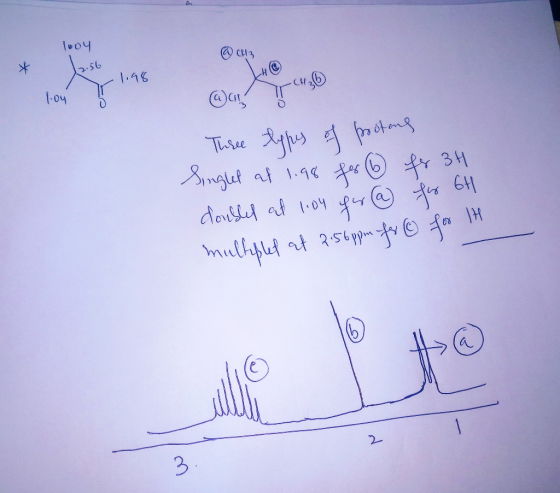
4. Draw a clear and accurate NMR spectrum for the molecule shown below. Clearly show the...
Part 2: Draw the NMR spectrum: Draw the structure of your molecule below, indicating each unique...
 Part 2: Draw the NMR spectrum: Draw the structure of your molecule below, indicating each unique hydrogen atom with letters, starting with A. After complete the below table, summarizing the integration, approximate chemical shift, number of neighbors, and splitting pattern of each hydrogen in your given molecule. Note, you do not need to fill every row in the below table. By CH₃ Structures H₂C- 2 Brono- 4 methyl pertane Hydrogen Integration Letter A Chemical Number of Splitting Pattern Shift Neighbors...
n-Butanol ОН Part 2: Draw the NMR spectrum Draw the structure of your molecule below, indicating...
 n-Butanol ОН Part 2: Draw the NMR spectrum Draw the structure of your molecule below, indicating each unique hydrogen atom with letters, starting with A. After complete the below table, summarizing the integration, approximate chemical shift number of neighbors, and splitting pattern of each hydrogen in your given molecule. Note, you do not need to fill every row in the below table. Structure: Hydrogen Integration Letter Chemical Number of Splitting Pattern Shift Neighbors Now, draw the NMR in the space...
Draw the expected H NMR spectrum for each molecule. Pay attention to the chemical shift and...
 Draw the expected H NMR spectrum for each molecule. Pay
attention to the chemical shift and splitting. Mark the integration
of each peak. Then assign each peak in the NMR to hydrogen atoms in
the structure.
HyC 0 ppm 192 t2 .3 +5 o t6 O=C +7 C=O +8
Draw the expected H NMR spectrum for each molecule. Pay
attention to the chemical shift and splitting. Mark the integration
of each peak. Then assign each peak in the NMR to hydrogen atoms in
the structure.
HyC 0 ppm 192 t2 .3 +5 o t6 O=C +7 C=O +8
Predicting the Spectrum MR and C NMR) the expected 'H NMR number of si spectrum for...
 Predicting the Spectrum MR and C NMR) the expected 'H NMR number of si spectrum for the following molecule. Be sure to include appropriate for the following molecule. Be sure to er of signals, show spisina ar try to indicate relative Integration by arca un (label peak with some splitting and try to indicate relative Integration by area under peak peak with correct number for clarity in tabel all the unique hydrogens with letters and assign them to the peaks...
PART 2 Draw the most accurate 'H-NMR spectrum you can for the following molecules. Show all...
 PART 2 Draw the most accurate 'H-NMR spectrum you can for the following molecules. Show all signals including an accurate representation of splitting, intensity and position. Assume no signal overlap (i.e. if you expect signals have similar chemical shifts, just place them next to each other). Clearly label the integration value for each signal. Indicate the correlation between each proton (HA, HB, etc) and each signal (label signals A, B, etc.) ppm 12.5 10 11 O ppm 12.5 10 ii...
3. Predict the 1H NMR spectrum of the molecule shown to the left. Be sure to include the x-axis for the 'H NMR spectrum...
 3. Predict the 1H NMR spectrum of the molecule shown to the left. Be sure to include the x-axis for the 'H NMR spectrum labelled with units, peaks for each of the non-equivalent protons clearly showing the expected splitting patterns, approximate chemical shifts, and integration
Draw the structure of the compound C3H6ICl that exhibits the NMR spectrum data below. Draw the...
 Draw the structure of the compound C3H6ICl that exhibits the NMR
spectrum data below.
Draw the structure of the compound CHIC that exhibits the from the DEPT data list. The triplet at 77 ppm is CDC.z. C-NMR spectrum and DEPT data below. Impurity peaks are omitted Flash Installation and Troubleshooting Shirt DEPT 20 DEPT 135 Used with permission from Aldrich Camical Co. Inc. Scent COC 35.4 25 positive peak negative par Chemical shift: 5 Y ZOOM 2250 1872 149.5 111.7...
Draw the structure of the compound C3H6ICl that exhibits the NMR
spectrum data below.
Draw the structure of the compound CHIC that exhibits the from the DEPT data list. The triplet at 77 ppm is CDC.z. C-NMR spectrum and DEPT data below. Impurity peaks are omitted Flash Installation and Troubleshooting Shirt DEPT 20 DEPT 135 Used with permission from Aldrich Camical Co. Inc. Scent COC 35.4 25 positive peak negative par Chemical shift: 5 Y ZOOM 2250 1872 149.5 111.7...
II. Number of Signals 2. Predict the number of unique protons that would generate peaks in an NMR spectrum. Look at...
 II. Number of Signals 2. Predict the number of unique protons that would generate peaks in an NMR spectrum. Look at Figure 13.6 and example problem 13.2 for guidance. Then for each of these unique proton signals, predict the ppm range where you would find it and the integration value or signal area. Look at figure 13.7 in your textbook. Finally, determine the splitting pattern (multiplicity) of the signal. This is predicted using the n+1 rule. Watch the video for...
Draw a H NMR spectrum for each compound. Pay attention to the chemical shift (ppm) multiplicity,...
 Draw a H NMR spectrum for each compound. Pay attention to the chemical shift (ppm) multiplicity, and integration of each peak. The boxes indicate where the peaks should go (which ppm range) as well as the relative height of each peak OH PPM O 3 PPM -2. H 10 8 PPM
 Part 2: Draw the NMR spectrum: Draw the structure of your molecule below, indicating each unique hydrogen atom with letters, starting with A. After complete the below table, summarizing the integration, approximate chemical shift, number of neighbors, and splitting pattern of each hydrogen in your given molecule. Note, you do not need to fill every row in the below table. By CH₃ Structures H₂C- 2 Brono- 4 methyl pertane Hydrogen Integration Letter A Chemical Number of Splitting Pattern Shift Neighbors...
 n-Butanol ОН Part 2: Draw the NMR spectrum Draw the structure of your molecule below, indicating each unique hydrogen atom with letters, starting with A. After complete the below table, summarizing the integration, approximate chemical shift number of neighbors, and splitting pattern of each hydrogen in your given molecule. Note, you do not need to fill every row in the below table. Structure: Hydrogen Integration Letter Chemical Number of Splitting Pattern Shift Neighbors Now, draw the NMR in the space...
 Draw the expected H NMR spectrum for each molecule. Pay
attention to the chemical shift and splitting. Mark the integration
of each peak. Then assign each peak in the NMR to hydrogen atoms in
the structure.
HyC 0 ppm 192 t2 .3 +5 o t6 O=C +7 C=O +8
Draw the expected H NMR spectrum for each molecule. Pay
attention to the chemical shift and splitting. Mark the integration
of each peak. Then assign each peak in the NMR to hydrogen atoms in
the structure.
HyC 0 ppm 192 t2 .3 +5 o t6 O=C +7 C=O +8
 Predicting the Spectrum MR and C NMR) the expected 'H NMR number of si spectrum for the following molecule. Be sure to include appropriate for the following molecule. Be sure to er of signals, show spisina ar try to indicate relative Integration by arca un (label peak with some splitting and try to indicate relative Integration by area under peak peak with correct number for clarity in tabel all the unique hydrogens with letters and assign them to the peaks...
 PART 2 Draw the most accurate 'H-NMR spectrum you can for the following molecules. Show all signals including an accurate representation of splitting, intensity and position. Assume no signal overlap (i.e. if you expect signals have similar chemical shifts, just place them next to each other). Clearly label the integration value for each signal. Indicate the correlation between each proton (HA, HB, etc) and each signal (label signals A, B, etc.) ppm 12.5 10 11 O ppm 12.5 10 ii...
 3. Predict the 1H NMR spectrum of the molecule shown to the left. Be sure to include the x-axis for the 'H NMR spectrum labelled with units, peaks for each of the non-equivalent protons clearly showing the expected splitting patterns, approximate chemical shifts, and integration
 Draw the structure of the compound C3H6ICl that exhibits the NMR
spectrum data below.
Draw the structure of the compound CHIC that exhibits the from the DEPT data list. The triplet at 77 ppm is CDC.z. C-NMR spectrum and DEPT data below. Impurity peaks are omitted Flash Installation and Troubleshooting Shirt DEPT 20 DEPT 135 Used with permission from Aldrich Camical Co. Inc. Scent COC 35.4 25 positive peak negative par Chemical shift: 5 Y ZOOM 2250 1872 149.5 111.7...
Draw the structure of the compound C3H6ICl that exhibits the NMR
spectrum data below.
Draw the structure of the compound CHIC that exhibits the from the DEPT data list. The triplet at 77 ppm is CDC.z. C-NMR spectrum and DEPT data below. Impurity peaks are omitted Flash Installation and Troubleshooting Shirt DEPT 20 DEPT 135 Used with permission from Aldrich Camical Co. Inc. Scent COC 35.4 25 positive peak negative par Chemical shift: 5 Y ZOOM 2250 1872 149.5 111.7...
 II. Number of Signals 2. Predict the number of unique protons that would generate peaks in an NMR spectrum. Look at Figure 13.6 and example problem 13.2 for guidance. Then for each of these unique proton signals, predict the ppm range where you would find it and the integration value or signal area. Look at figure 13.7 in your textbook. Finally, determine the splitting pattern (multiplicity) of the signal. This is predicted using the n+1 rule. Watch the video for...
 II. Number of Signals 2. Predict the number of unique protons that would generate peaks in an NMR spectrum. Look at Figure 13.6 and example problem 13.2 for guidance. Then for each of these unique proton signals, predict the ppm range where you would find it and the integration value or signal area. Look at figure 13.7 in your textbook. Finally, determine the splitting pattern (multiplicity) of the signal. This is predicted using the n+1 rule. Watch the video for...
 Draw a H NMR spectrum for each compound. Pay attention to the chemical shift (ppm) multiplicity, and integration of each peak. The boxes indicate where the peaks should go (which ppm range) as well as the relative height of each peak OH PPM O 3 PPM -2. H 10 8 PPM
Most questions answered within 3 hours.
-
Calculate the number density of argon gas at a temperature of
24C and a pressure of...
asked 2 hours ago -
Alternative
Classification
How to Estimate
Probabilities from Data? ( For continuous Attributes)
And How to generate...
asked 2 hours ago -
An explosion breaks a 20.0-kg object into three parts. The
object is initially moving at a...
asked 3 hours ago -
Calculate the approximate number of residues of Rubisco, which
is involved in carbon fixation in plants,...
asked 4 hours ago -
Other decisions about scientific claims can have a much broader
impact.ENERGYarrow-10x10.png, environment, health, security - all...
asked 5 hours ago -
I need to write a research paper and work cited about this
topic: The United States...
asked 5 hours ago -
Hello! I was wondering if I could have some help?
If the vapor pressure of carvone...
asked 6 hours ago -
An economist wants to estimate the mean per capita income (in
thousands of dollars) for a...
asked 6 hours ago -
What would be the input/output characteristic of a circuit
obtained by putting two of your 2's-complementers...
asked 6 hours ago -
In Drosophila, the transition from the syncytial blastoderm
stage to the cellular blastoderm stage is a...
asked 7 hours ago -
Project management question:
Name 3 different types of resources (hint: humans are one
type)
asked 7 hours ago -
Consider the following reaction: C 2H 2( g) + 2H 2( g) C 2H 6(
g)...
asked 7 hours ago