- List 3 different molecular methods that could be used to detect the 1p/19q deletion. Describe what would be seen for each method if 1p/19q co-deletion were present. NOTE: LIST ONLY MOLECULAR METHODS SUCH AS FISH AND PCR AND DESCRIBE WHAT WOULD BE SEEN.
- The cells of oligodengroglioma do not grow well in culture. Of the three assays from question 2, which would you choose to diagnose the 1p/19q deletion? Why? AGAIN LIST ONE OF THE MOLECULAR METHODS, NO OTHER METHODS ARE ACCEPTABLE!!
Homework Answers
Ans) The 1p 19q deletion status can be analyzed with various molecular-genetic methods including FISH, comparative genomic hybridization (CGH), chromogenic in situ hybridization (CISH), PCR-based microsatellite analysis, real-time comparative quantitative PCR and multiplex ligation-dependent probe amplification (MLPA)
- Complete deletion of both the short arm of chromosome 1 (1p) and the long arm of chromosome 19 (19q) (1p/19q co-deletion) is the molecular genetic signature of oligodendrogliomas, a subtype of primary brain tumours accounting for approximately ten to fifteen percent of all diffuse gliomas in adults. The loss of one hybrid chromosome results in 1p and 19q loss of heterozygosity (LOH) 3. This molecular alteration is the result of an unbalanced whole-arm translocation between chromosomes 1 and 19 3 with the loss of the derivative t(1p;19q), which occurs early in the pathogenesis of oligodendrogliomas. Initially described in 1994 4, the biologic effect of 1p/19q co-deletion remains unclear. 1p/19q co-deletion is a valuable diagnostic, prognostic and predictive biomarker for the management of oligodendroglial tumours.
- 1p/19q co-deletion as a diagnostic biomarker in glioma
1p/19q co-deletion is a pathognomonic biomarker that defines a
distinct glioma entity 5 and is characteristic of
oligodendrogliomas 6,7. Virtually, all 1p/19q co-deleted
oligodendrogliomas have mutation in isocitrate dehydrogenase 1
(IDH1) at arginine 132 (R132) or the analogous residue arginine 172
in IDH2 (R172). Other common molecular alterations co-occuring with
1p/19q co-deletion include mutations in the telomerase reverse
transcriptase (TERT) gene promoter, mutations in homolog of
Drosophila capicua (CIC) and far upstream element binding protein
(FUBP1) 9, and promoter methylation of the methyl-guanine methyl
transferase (MGMT) gene 8,7. With very few exceptions, 1p/19q
co-deletion is mutually exclusive with TP53 and ATRX mutation,
which both characterize glial tumours of astrocytic lineage.
- Thereby, assessment of 1p/19q co-deletion, together with IDH mutation status and other molecular markers (e.g. ATRX and TP53 status), can help distinguish oligodendrogliomas which are IDH-mutant and 1p/19q-codeleted, from tumours of astrocytic lineage which are 1p/19q-non co-deleted.
- In some cases, an oligodendroglioma can be cured. Anaplastic oligodendroglioma (grade III): An anaplastic oligodendroglioma grows quickly and spreads into nearby tissues. The tumor cells look different from normal cells. This type of tumor usually cannot be cured.
Add Answer to:
List 3 different molecular methods that could be used to detect
the 1p/19q deletion. Describe what...
Explain the results in the Methylation Specific PCR (MSP) above. Be specific The positive control in...
 Explain the results in the Methylation Specific PCR (MSP)
above. Be specific
The positive control in the lower panel is much stronger than
the patient samples. What control would be better (or in addition
to this control) to detect the weak expression in T3 and T4? Be
specific
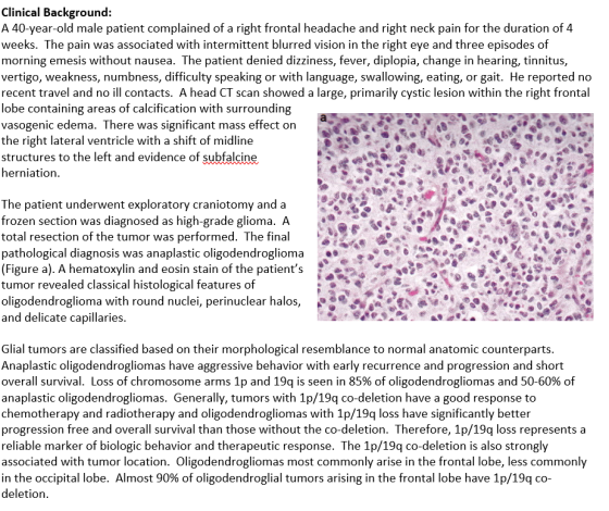
Clinical Background: A 40-year-old male patient complained of a right frontal headache and right neck pain for the duration of 4 weeks. The pain was associated with intermittent blurred vision in the...
Explain the results in the Methylation Specific PCR (MSP)
above. Be specific
The positive control in the lower panel is much stronger than
the patient samples. What control would be better (or in addition
to this control) to detect the weak expression in T3 and T4? Be
specific
Clinical Background: A 40-year-old male patient complained of a right frontal headache and right neck pain for the duration of 4 weeks. The pain was associated with intermittent blurred vision in the...
 Explain the results in the Methylation Specific PCR (MSP)
above. Be specific
The positive control in the lower panel is much stronger than
the patient samples. What control would be better (or in addition
to this control) to detect the weak expression in T3 and T4? Be
specific
Clinical Background: A 40-year-old male patient complained of a right frontal headache and right neck pain for the duration of 4 weeks. The pain was associated with intermittent blurred vision in the...
Explain the results in the Methylation Specific PCR (MSP)
above. Be specific
The positive control in the lower panel is much stronger than
the patient samples. What control would be better (or in addition
to this control) to detect the weak expression in T3 and T4? Be
specific
Clinical Background: A 40-year-old male patient complained of a right frontal headache and right neck pain for the duration of 4 weeks. The pain was associated with intermittent blurred vision in the...
Most questions answered within 3 hours.
-
Please help me with FLOWCHART and UML diagram for class,
thank you!
#include <iostream>
#include <fstream>...
asked 16 minutes ago -
3. Describe the “logic circuit” of the Lac operon. Which
proteins are bound or not to...
asked 17 minutes ago -
Ayesha’s adjusted gross income is $60,000 in 2019. She donated a
piece of artwork with a...
asked 24 minutes ago -
For Dijkstra’s shortest path algorithm:
a. Give the Big-O time for Dijkstra’s shortest path algorithm
and...
asked 36 minutes ago -
Phosphorus violates the 'octet rule' in biological molecules,
forming more covalent bonds than expected based on...
asked 39 minutes ago -
A 1.3 eV electron has a 10-4 probability of tunneling
through a 2.4 eV potential barrier....
asked 57 minutes ago -
What is the one ingredient that is common to being successful
with all stakeholders?
profit
trust...
asked 56 minutes ago -
Write an assembly language 32 bit program that reads in lines of
text by a .txt...
asked 59 minutes ago -
what is the density ( in g/L) of hydrogen gas at 29 degrees C and a...
asked 1 hour ago -
5-6. You are considering three investment alternatives for some
spare cash: Old Reliable Corporation stock (A1),...
asked 59 minutes ago -
Problem 16-02
Receivables Investment
Medwig Corporation has a DSO of 45 days. The company averages
$7,250...
asked 1 hour ago -
Mr. Brown hired Lowe's Maintenance Services Limited to repair
and paint the exterior wall of his...
asked 1 hour ago