1.


2.


4.Thr answers are already given in question no 1.
AUTONOMIC NERVOUS SYSTEM IS DIVIDED INTO (FIG. 87.11 A. The parasympathetic division B. The sympathetic division. The parasympathetic division The parasympathetic efferents come out as craniosacral outflow: 1. The cranial outflow: a. It is the part of the parasympathetic system coming out from the brain via 3rd (oculomotor), 7th (facial), 9th (glossopharyngeal) and 10th (vagus) nerves. b. The cell bodies of the preganglionic neurons are located in the cor- responding cranial nerve nuclei within the brain. C. The postganglionic neurons arise from ganglia usually close to the target organ. The ciliary ganglion, the otic ganglion are examples. 2. The sacral outflow: a. The sacral outflow arises from the ermediolateral gray column or lateral horn of 2nd to 4th sac segments. b. The cell bodies of the pregang neurons are located in the la preganglionic
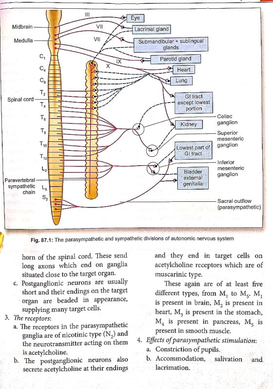
- Midbrain- VII Medulla Eye Lacrimal gland Submandibular + sublingual glands Parotid gland Heart Lung XI X 111 o or UTILILIT TIILIT Spinal cord GI tract except lowest portion علاماه Celiac ganglion Kidney Superior mesenteric ganglion Lowest part of Gl tract TOT Thanam Inferior mesenteric ganglion jo Paravertebral – sympathetic chain Bladder external genitalia -Sacral outflow (parasympathetic) Fig. 87.1: The parasympathetic and sympathetic divisions of autonomic nervous system horn of the spinal cord. These send long axons which end on ganglia situated close to the target organ. c. Postganglionic neurons are usually short and their endings on the target organ are beaded in appearance, supplying many target cells. 3. The receptors: a. The receptors in the parasympathetic ganglia are of nicotinic type (N,) and the neurotransmitter acting on them is acetylcholine. b. The postganglionic neurons also neurons also secrete acetylcholine at their endings and they end in target cells on acetylcholine receptors which are of muscarinic type. These again are of at least five different types, from M, to Ms. M, is present in brain, M, is present in heart, M, is present in the stomach, MA is present in pancreas, M, is present in smooth muscle. 4. Effects of parasympathetic stimulation: a. Constriction of pupils. b. Accommodation, salivation and l acrimation.
ITILIPILJULJIVIU9) the ganglia and rela e supplied to differem ich the postganglionic spinal nerves communicantes the spinal nerves nic effectors such as onic efferents may c. Negative inotropic, chronotropic, dromotropic and bathmotropic effect on heart. d. Contraction of gastrointestinal, geni- tourinary and respiratory muscles but relaxation of sphincters. . e. Secretion of glands, e.g. gastric, pancreatic, lacrimal, bronchial, etc. The sympathetic division 1. The sympathetic efferents come out from the spinal cord as thoracolumbar outflow which extends from the 1st thoracic segment (T) to the 3rd or 4th lumbar segments (L/L.). 2. The preganglionic efferent neurons arise from cell bodies located in the lateral horn of the spinal cord (intermediolateral gray column). 3. The preganglionic efferents come out through the anterior (ventral) root of the spinal segment and then pass through the white rami communicantes (which connect the ventral root to the paravertebral sympathetic chain) to the paravertebral sympathetic chain. (Fig. 87.2). Here, most of the preganglionic neurons end at the ga with postganglionic neuron 4. Postganglionic neurons: a. They may be suppli viscera on which th nerves end, b. They may return to the spinal through gray rami commi and spread along the spinal to supply autonomic effector vascular smooth muscles. c. Some preganglionic efferent pass right through the parave ganglion chain without interru They end in some collateral da located close to the viscera. (Th are the celiac, superior mesenteris and inferior mesenteric ganglia) From there, postganglionic neurons supply the viscera. d. The male and female reproductive organs are in part supplied by a special type of postganglionic sympathetic innervation. Here, the sympathetic postganglionic neurons are very short, the ganglia lying very close to or on the viscera. hout interruption. Dorsal root Paravertebral sympathetic chain Preganglionic sympathetic neuron Gray ramus communicantes White ramus communicantes Postganglionic sympathetic neuron supplying blood vessels, sweat glands, etc. Fig. 87.2: Sympathetic preganglionic and postganglionic neurons running white and gray ramus communicantes respectively cheurons running through
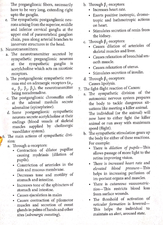
The preganglionic fibers, necessarily have to be very long, extending right upto the ganglia. e. The sympathetic postganglionic neu- rons arising from the superior, middle and inferior cervical ganglia at the upper end of paravertebral ganglion chain, pass along arterial branches to innervate structures in the head. 5. Neurotransmitters: a. The neurotransmitter secreted by sympathetic preganglionic neurons at the sympathetic ganglia is acetylcholine which acts on nicotinic receptors. b. The postganglionic sympathetic neu rons end on adrenergic receptors (ag, a., B, B, B2), the neurotransmitter being noradrenaline. c. The postganglionic chromaffin cells at the adrenal medulla secrete adrenaline (epinephrine). d. Some postganglionic sympathetic neurons secrete acetylcholine at their endings (blood vessels of skeletal muscles supplied by cholinergic vasodilator system). 6. The main actions of sympathetic divi- sion: a. Through a-receptors: • Contraction of dilator pupillae causing mydriasis (dilation of pupils). Constriction of arterioles in the skin and mucous membrane. • Decreases tone and motility of stomach and intestine. • Increases tone of the sphincters of stomach and intestine. Causes ejaculation in males Causes contraction of pilomotor muscles and secretion of sweat glands in palms of hands and other sites (adrenergic sweating). b. Through B, -receptors: Increases heart rate. Exerts positive inotropic, dromo- tropic and bathmotropic actions on heart. Stimulates secretion of renin from the kidney. c. Through B -receptors: • Causes dilation of arterioles of skeletal muscles and liver. . Causes relaxation of bronchial sm- ooth muscle. Causes relaxation of uterus. • Stimulates secretion of insulin. . d . Through B,-receptors: Causes lipolysis. 7. The fight-flight reaction of Canon: a. The sympathetic division of the autonomic nervous system prepares the body to tackle dangerous sit- uations like meeting a killer animal. The individual (or the animal) will now have to either fight the killer animal or run away with maximum speed (flight) b. The sympathetic stimulation gears up the body for either of these reactions. For example: • There is dilation of pupils—This allows passage of more light to the retina improving vision. • There is increased heart rate and elevated blood pressure—This helps in increasing perfusion of im-portant organs and muscles. There is cutaneous vasoconstric- tion-This restricts blood loss from surface wounds. • The threshold of activation of reticular formation is lowered- This helps the individual to maintain an alert, aroused state.
There is increased lipolysis and glycogenolysis—This provides ad- ditional nutrients and energy. c. Thus, it may be observed that all the effects of sympathetic stimulation help the individual or animal in meeting the emergency situation. In other words, the sympathetic nervous system is well-equipped to meet the immediate needs of an emergency. For this reason, the sympathetic nerve is also known as 'nerve of today'. d. In contrast, the parasympathetic division of the autonomic nervous system decreases heart rate, helps in digestion and absorption of food by increasing gut motility and secretion of glands. Thus, it helps in the day-to-day vegetative activity and conserves energy. So, it is called 'nerve of tomorrow'. to three ganglia called superior, middle and inferior cervical ganglia which form the topmost part of the paravertebral sympathetic ganglionic chain. 2. From these ganglia, the postganglionic neurons emerge and form a plexus around carotid artery and reach different organs along its branches. 3. Horner's syndrome is caused by a lesion anywhere in the course of these sym- pathetic neurons. Thus, the site of lesion may be preganglionic, at the level of the ganglia or postganglionic. | Horner's syndrome results in: a. Miosis (constriction of pupil)-due to paralysis of dilator pupillae. b. Enophthalmos (sunken eyes)—due to | paralysis of orbitalis muscle. c. Anhidrosis of one-half of the face- absence of sweating due to damage to sympathetic sudomotor fibers. d. Ptosis—due to paralysis of Müller's muscle. e. Loss of citiospinal reflex. (Ciliospinal reflex-dilation of pupils | on scratching the skin of the neck.] APPLIED Horner's syndrome: 1. The sympathetic nerve fibers supplying the head and neck region arise from the T, and T, segments and then pass
Acetylcholine: 1. It is the neurotransmitter secreted at the preganglionic nerve-endings of both sympathetic and parasympathetic nervous system, at the postganglionic parasympathetic nerve-endings, also at the neuromuscular junction and many CNS synapses. 2. Its receptor is principally of two types: (a) Nicotinic and (b) Muscarinic. 3. Nicotinic cholinergic receptors are present in: (a) The autonomic ganglia, (b) in neuromuscular junction and (c) in CNS. [a-bungarotoxin blocks the N, nicotinic receptors at neuromuscular junction but has no effect at the ganglia. On the other hand, the drug mecamylamine blocks N, nicotinic receptors at the ganglia but has no effect on the N, receptors at the neuromuscular junction.] 4. Nicotinic cholinergic receptors are formed by 5 subunits which encircle a central ion channel. The channel remains closed in the inactive state. The composition of the subunits varies with the type and location of the receptor but usually contains 2a, ß, y and d subunits. 5. When acetylcholine becomes attached to the binding sites present on a-subunits of the receptor, a conformational change occurs, which results in opening of the
4. Both a and B-receptors are serpentine receptors coupled with G proteins. DOPAMINE channel allowing passage of Nat and other small ions. This results in the formation of EPSP or EPP as the case may be, and if threshold is reached, this will result in the formation of an action potential. 6. The muscarinic receptors are of different varieties. They are of five types-M, to M. Mostly, they are serpentine receptors and act through G proteins. 7. M, type of receptor is present in brain. M, is found in heart, M, is present in pancreas, mediating both exocrine and endocrine function while M, and M. mediate smooth muscle activity. 8. Atropine blocks all types of muscarinic receptors. The drug pirenzepine is a selective M -blocker blocking receptors in oxyntic cells which secrete HCI. Hence, it is used in the treatment of peptic ulcer, I NOREPINEPHRINE AND EPINEPHRINE 1. This is another catecholamine neuro- transmitter present in the synaptic junctions at certain areas of the brain. 2. Dopamine receptors are mainly of 5 types, D, to Ds 3. Dopamine, as a neurotransmitter, activates two different dopaminergic systems. One is called the nigrostriatal dopamine system and the other is called mesolimbic dopamine system. 4. The nigrostriatal system extends from substantia nigra in the midbrain to corpus striatum in the basal ganglia. Degeneration of this system produces Parkinson's disease (see p. 713). Dopaminergic drugs like L-dopa which restore the deficient dopamine, are useful in treating this disease. 5. The mesolimbic dopamine system extends from the midbrain to limbic area of the forebrain. Overactivity of this system has effects on mood and beha- vior and is also implicated in addiction to substances like amphetamine, cocaine, alcohol and even nicotine. Imbalance of dopamine in this system may cause schizophrenia-A disease involving mood and behavior. This disease is treated by D,-blocker drugs and a pre- dictable side-effect is symptoms like Parkinson's disease. 6. Newer 'atypical antipsychotics have minimal extrapyramidal side-effects as they have less activity on striatal D,- receptors. 1. These are catecholamine neurotrans- mitters. Both are secreted from the adrenal medulla but only norepinephrine is secreted from sympathetic post-ga- nglionic nerve-endings. Epinephrine-secreting neurons are present in the brain. (Synthesis, metabolism and actions of these catecholamines have been discussed in connection with adrenal medulla; see p. 561). 2. Both norepinephrine and epinephrine act on a and B-receptors. a-receptors may be of a, and a, types. B-receptors are classified into B B, and ß3 types. 3. Norepinephrine has more pronounced effect on a-receptors while epinephrine has a predominant effect on B-receptors.
I SEROTONIN regulation of blood pressure, water intake, pain threshold, sexual behavior, etc. 4. H, -antagonists, in addition to antihistaminic action, produce sedation and weight gain on chronic use. The drowsiness produced by conventional antiallergic drugs are known to all. GLUTAMATE AND ASPARTATE 1. Serotonin, which is derived from the amino acid tryptophan, is another important neurotransmitter. 2. It is found in the platelets, in the brain and in the enterochromaffin cells and myenteric plexus in the gastrointestinal tract. 3. Serotonin has numerous receptors. These are classified as 5HT, to 5HT, Again, 5HT, 5HT, and 5HT, groups are further subclassified. For example, 5HT, group contains 5HT,4,5HT, and 5HT,c types of receptors. 4. Newer drugs have been evolved with action on specific 5HT (serotonin) receptors. For example, the drug sumatriptan relieves migraine by its selective agonist action on 5HT, receptor, the drug ondansetron relieves nausea and vomiting by its antagonist action on 5HT, receptor, the drug mosapride is a gastroprokinetic and it prevents gastroesophageal reflux by its selective agonist action on 5HTA receptor. 5. Use of SSRI (selective serotonin reuptake inhibitor) drugs has been mentioned above. 1. These are excitatory neurotransmitters. Glutamate is involved in 75% of all excitatory transmission in the brain. 2. Glutamate has two types of receptors: a. Metabotropic—These are coupled to G-proteins. b. lonotropic-Like nicotinic choliner- gic receptors, these are ion channels. There are 3 ionotropic receptors Kainate, AMPA and NMDA. 3. NMDA (N-Methyl D-Aspartate) re- ceptors: a. These are involved in long-term potentiation, learning and memory. [In longterm potentiation, a high frequency stimulation for a short duration facilitates transmission of impulses across synapses for a prolonged period.] b. NMDA receptors are abundant in hippocampus. They have following special features: • They require glycine for activation. A Mg++ ion keeps the channel blocked at resting membrane potential even if glutamate binds to it. However, if the neuron is depolarized through a separate glutamate-sensitive AMPA recep- tor, the Mg++-block is removed. IHISTAMINE 1. Histamine is synthesized by de- carboxylation of the amino acid histidine. As a neurotransmitter, it is distributed widely in brain and spinal cord, stomach and the pituitary gland. 2. There are three types of receptors, H, H, and Hz. H, receptor is mainly presynaptic. 3. Histamine is believed to mediate a wide range of CNS functions, such as,
UU IMDA receptor ver, it has also ion mediated glycine is proved uscle excitability • Now, Cat+ ions enter in large 5. Glycine: a. When it binds to NMDA amounts into the neuron. In long- it is excitatory. However, it ha term potentiation, this ultimately a direct inhibitory action medi leads to: like GABA, by a Cl channel. - Augmented activity of AMPA b. Inhibitory action of glycine is n receptors. by the increased muscle excita - Migration of more AMPA and seizures produced by strych receptors to the postsynaptic a glycine antagonist. membrane. OPIOID PEPTIDES Additionally, a retrograde signal in the form of NO or arachidonate 1. The drug morphine binds to receptors is is believed to pass from the the brain and gastrointestinal tract. These postsynaptic to the presynaptic receptors are called opioid receptors. neuron, increasing further release 2. Investigations were carried out with of neurotransmitter. a view to finding out the body's own • All these events result in long-term ligands for the morphine-binding potentiation, which, in turn, is receptors or opioid receptors. As a result involved in learning and memory. of the search, several peptides have been discovered, which are called 'opioid I GABA AND GLYCINE peptides'. 1. GABA and glycine are inhibitory 3. These opioid peptides are: (a) Enc- neurotransmitters. GABA (v-amino ephalins, (b) endorphins and (c) butyric acid) is widely present in the dynorphins. aynorphins. brain. It is formed by decarboxylation of of 4. Encephalins include met-encephalin 9 glutamate. which contains methionine and leu- 2. There are 3 GABA receptors, GABA encephalin which contains leucine. GABAR and GABAC GABA, and 5. The opioid peptides are derived from GABA, are Cl- channels formed by five three main precursors: subunits encircling a small pore like a. Proencephalin-Which gives rise the nicotininic cholinergic receptor. to leu-encephalin, met-encephalin, In contrast, GABA, is coupled to G octapeptide and a heptapeptide. proteins. b. Pro-opiomelanocortin-Which gives 3. Cl- influx through Cl-channels leads to rise to B-endorphin and other end hyperpolarization of the postsynaptic orphins. neuron with formation of IPSP. c. Prodynorphin-Which gives rise 4. Benzodiazepine, a widely used drug with dynorphins and neoendorphins. 6. The opioid peptides have the anxiolytic and sedative action, binds to the a-subunit of GABA, receptor and receptors H, K and 8. They are cous to G proteins of Gq type and de increases Cl- conductance. CAMP. des have three types of Y are coupled ype and decrease


 Create a map or outline of the nervous system and include the
required data:
Divisions of the nervous system and function
Neurotransmitters of the Sympathetic/Parasympathetic Nervous
System
Sympathetic Response/Parasympathetic Response
Example of Adrenergic Rx & It's therapeutic use
Example of Adrenergic Blocker Rx & It's therapeutic
use
Example of Cholinergic Rx & It's therapeutic use
Example of Cholinergic Blocker Rx & therapeutic use
NERVOUS SYSTEM Peripheral Nervous System CENTRAL NERVOUS SYSTEM Consists of: Brain and spinal cord All the rest...
Create a map or outline of the nervous system and include the
required data:
Divisions of the nervous system and function
Neurotransmitters of the Sympathetic/Parasympathetic Nervous
System
Sympathetic Response/Parasympathetic Response
Example of Adrenergic Rx & It's therapeutic use
Example of Adrenergic Blocker Rx & It's therapeutic
use
Example of Cholinergic Rx & It's therapeutic use
Example of Cholinergic Blocker Rx & therapeutic use
NERVOUS SYSTEM Peripheral Nervous System CENTRAL NERVOUS SYSTEM Consists of: Brain and spinal cord All the rest...