
Homework Answers
Add Answer to:
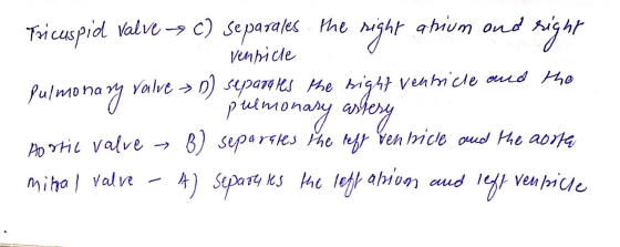
Check Your Understanding 1.1 Match the heart valve with the structures separated by each valve. Tricuspid...
Pulmonary semilunar valve Right atrium Left atrium Left AV (mitral or bicuspid) valve Right AV (tricuspid)...
 Pulmonary semilunar valve Right atrium Left atrium Left AV (mitral or bicuspid) valve Right AV (tricuspid) valve Left pulmonary artery (to left lung) Right pulmonary artery (to right lung) Right pulmonary veins (from right lung) Inferior vena cava Loft pulmonary veins (from left lung) Superior vena cava (from upper body) Left ventricle Chordae tondinese Interventricular septum Papillary muscle Pulmonary trunk Descending aorta (to lower body) Right ventricle Aortic semilunar valve Aorta (to systemic organs)
Anatomy 9. Starting with the right atrium, trace a drop of blood through the heart and...
 Anatomy
9. Starting with the right atrium, trace a drop of blood through the heart and lungs, naming the following structures: aorta, aortic valve, left atrium, left ventricle, mitral valve, pulmonary arteries, pulmonary capillaries, pulmonary valve, pulmonary trunk, pulmonary veins, right atrium, right ventricle, and tricuspid valve.
Anatomy
9. Starting with the right atrium, trace a drop of blood through the heart and lungs, naming the following structures: aorta, aortic valve, left atrium, left ventricle, mitral valve, pulmonary arteries, pulmonary capillaries, pulmonary valve, pulmonary trunk, pulmonary veins, right atrium, right ventricle, and tricuspid valve.
HEART CHAMBERS. MATCH LETTER WITH NUMBER 1.sends blood to the tricuspid valve 2.sends blood...
HEART CHAMBERS. MATCH LETTER WITH NUMBER 1.sends blood to the tricuspid valve 2.sends blood to the pulmonary semilunar valve 3.sends blood to the bicuspid valve 4.sends blood to the aortic semilunar valve 5.upper chambers 6.lower chambers 7.carry deoxygenated blood 8.carry oxygenated blood 9.blood entering here has come from the pulmonary arteries 10.blood entering here has come from the pulmonary veins A. BOTH Left Atrium and Left Ventricle B. Right Atrium...
Name: A. Match the following terms with their meanings below: superior vena cava. tricuspid valve ventri...
 Name: A. Match the following terms with their meanings below: superior vena cava. tricuspid valve ventricle aorta mitral valve arteriole atrium pulmonary artery pulmonary vein capillary 1. Smallest blood vessel. 2. Largest artery in the body 3. Lower chamber of the heart 4. Valve between the right atrium and ventricle 5. Carries blood from the lungs to the heart 6. Brings blood to heart from upper parts of the body 7. Upper chamber of the heart 8. Valve between the...
Matching: 34. tricuspid valve epicardium bicuspid valve myocardium chordea tendinea semilunar valve endocardium A. aortic and...
 Matching: 34. tricuspid valve epicardium bicuspid valve myocardium chordea tendinea semilunar valve endocardium A. aortic and pulmonary valve B. valve between right atrium and right ventricle C. middle layer of the heart, cardiac muscle tissue D. attached to bicuspid and tricuspid valves E. valve between left atrium and left ventricle F. deepest layer of the heart, lines heart chambers G. outer layer of the heart, also called visceral pericardium
7. (6 points) Matching: Match each term with the correct definition. A. Located between the left...
 7. (6 points) Matching: Match each term with the correct definition. A. Located between the left ventricle and the aorta B. Located between the left atrium and left ventricle 1. Myocardium 2. Parietal pericardium 3. Tricuspid valve 4. Aortic valve 5. Papillary muscles 6. Pulmonary veins F. Bring(s) deoxygenated blood to the lungs 7. Mitral valve 8. Visceral pericardium H. Vein of the coronary circulation 9. Pulmonary trunk l. Outer layer of the serous pericardium 10. Coronary sinusJ. Thread-like structure(s)...
Identify the components of the intrinsic conduction system Matching: tricuspid valve epicardium bicuspid valve myocardium chordea...
 Identify the components of the intrinsic conduction system Matching: tricuspid valve epicardium bicuspid valve myocardium chordea tendinea semilunar valve endocardium A aortic and pulmonary valve B. valve between right atrium and right ventricle C. middle layer of the heart, cardiac muscle tissue D. attached to bicuspid and tricuspid valves E valve between left atrium and left ventricle F. deepest layer of the heart, lines heart chambers G. outer layer of the heart, also called visceral pericardium I Forum
Label the structures indicated on this anterior view of the Internal anatomy of the heart model....
 Label the structures indicated on this anterior view of the Internal anatomy of the heart model. Left ventricle 0.37 points Right ventricle Skipped Left atrium References Pulmonary semilunar valve Lolt AV Valve (blouspid or mitral) Right atrium Right AV valve (tricuspid) Zoom Reset
ANATOMY Instructions say Start with SUPERIOR MESENTERIC VEIN & END WITH SUPERIOR MESENTERIC ARTERY I provided...
 ANATOMY Instructions say
Start with SUPERIOR MESENTERIC VEIN &
END WITH SUPERIOR MESENTERIC ARTERY
I provided and example of how they want this done , example is
the one showing the little arrows etc . Thank you
They want arrows not paragraphs .
Appreciate your time & help
Followin Following is an example in which we have started in the right popliteal vein and ended in ave started in the right popliteal vein and ende d in the left internal...
ANATOMY Instructions say
Start with SUPERIOR MESENTERIC VEIN &
END WITH SUPERIOR MESENTERIC ARTERY
I provided and example of how they want this done , example is
the one showing the little arrows etc . Thank you
They want arrows not paragraphs .
Appreciate your time & help
Followin Following is an example in which we have started in the right popliteal vein and ended in ave started in the right popliteal vein and ende d in the left internal...
 Pulmonary semilunar valve Right atrium Left atrium Left AV (mitral or bicuspid) valve Right AV (tricuspid) valve Left pulmonary artery (to left lung) Right pulmonary artery (to right lung) Right pulmonary veins (from right lung) Inferior vena cava Loft pulmonary veins (from left lung) Superior vena cava (from upper body) Left ventricle Chordae tondinese Interventricular septum Papillary muscle Pulmonary trunk Descending aorta (to lower body) Right ventricle Aortic semilunar valve Aorta (to systemic organs)
 Anatomy
9. Starting with the right atrium, trace a drop of blood through the heart and lungs, naming the following structures: aorta, aortic valve, left atrium, left ventricle, mitral valve, pulmonary arteries, pulmonary capillaries, pulmonary valve, pulmonary trunk, pulmonary veins, right atrium, right ventricle, and tricuspid valve.
Anatomy
9. Starting with the right atrium, trace a drop of blood through the heart and lungs, naming the following structures: aorta, aortic valve, left atrium, left ventricle, mitral valve, pulmonary arteries, pulmonary capillaries, pulmonary valve, pulmonary trunk, pulmonary veins, right atrium, right ventricle, and tricuspid valve.
 Name: A. Match the following terms with their meanings below: superior vena cava. tricuspid valve ventricle aorta mitral valve arteriole atrium pulmonary artery pulmonary vein capillary 1. Smallest blood vessel. 2. Largest artery in the body 3. Lower chamber of the heart 4. Valve between the right atrium and ventricle 5. Carries blood from the lungs to the heart 6. Brings blood to heart from upper parts of the body 7. Upper chamber of the heart 8. Valve between the...
 Matching: 34. tricuspid valve epicardium bicuspid valve myocardium chordea tendinea semilunar valve endocardium A. aortic and pulmonary valve B. valve between right atrium and right ventricle C. middle layer of the heart, cardiac muscle tissue D. attached to bicuspid and tricuspid valves E. valve between left atrium and left ventricle F. deepest layer of the heart, lines heart chambers G. outer layer of the heart, also called visceral pericardium
 7. (6 points) Matching: Match each term with the correct definition. A. Located between the left ventricle and the aorta B. Located between the left atrium and left ventricle 1. Myocardium 2. Parietal pericardium 3. Tricuspid valve 4. Aortic valve 5. Papillary muscles 6. Pulmonary veins F. Bring(s) deoxygenated blood to the lungs 7. Mitral valve 8. Visceral pericardium H. Vein of the coronary circulation 9. Pulmonary trunk l. Outer layer of the serous pericardium 10. Coronary sinusJ. Thread-like structure(s)...
 Identify the components of the intrinsic conduction system Matching: tricuspid valve epicardium bicuspid valve myocardium chordea tendinea semilunar valve endocardium A aortic and pulmonary valve B. valve between right atrium and right ventricle C. middle layer of the heart, cardiac muscle tissue D. attached to bicuspid and tricuspid valves E valve between left atrium and left ventricle F. deepest layer of the heart, lines heart chambers G. outer layer of the heart, also called visceral pericardium I Forum
 Label the structures indicated on this anterior view of the Internal anatomy of the heart model. Left ventricle 0.37 points Right ventricle Skipped Left atrium References Pulmonary semilunar valve Lolt AV Valve (blouspid or mitral) Right atrium Right AV valve (tricuspid) Zoom Reset
 Label the structures indicated on this anterior view of the Internal anatomy of the heart model. Left ventricle 0.37 points Right ventricle Skipped Left atrium References Pulmonary semilunar valve Lolt AV Valve (blouspid or mitral) Right atrium Right AV valve (tricuspid) Zoom Reset
 ANATOMY Instructions say
Start with SUPERIOR MESENTERIC VEIN &
END WITH SUPERIOR MESENTERIC ARTERY
I provided and example of how they want this done , example is
the one showing the little arrows etc . Thank you
They want arrows not paragraphs .
Appreciate your time & help
Followin Following is an example in which we have started in the right popliteal vein and ended in ave started in the right popliteal vein and ende d in the left internal...
ANATOMY Instructions say
Start with SUPERIOR MESENTERIC VEIN &
END WITH SUPERIOR MESENTERIC ARTERY
I provided and example of how they want this done , example is
the one showing the little arrows etc . Thank you
They want arrows not paragraphs .
Appreciate your time & help
Followin Following is an example in which we have started in the right popliteal vein and ended in ave started in the right popliteal vein and ende d in the left internal...
Most questions answered within 3 hours.
-
Write a program to solve the Josephus problem, with the following
modification:
Sample Input:
./a.out n...
asked 8 minutes ago -
At the start of a CD it is spinning at a rate of 525 rpm
(revolutions...
asked 44 minutes ago -
4. Without doing any calculations, predict whether the observed
∆T would increase, decrease or remain the...
asked 1 hour ago -
Based on the range, which of the following sets of scores has
the greatest variability? 3,...
asked 3 hours ago -
Ripples in a pond travel at a velocity of 3 m/s with one peak
passing a...
asked 2 hours ago -
A man stands on the roof of a building of height 13.0 mm and
throws a...
asked 3 hours ago -
The extent to which assets are financed by borrowed funds and
other liabilities is indicated by:...
asked 4 hours ago -
Explain in detail
Germany is the fifth largest economy
explain what goods and services Germany specializes...
asked 4 hours ago -
The density of platinum is 21.45 g/mL. If a cube of platinum
with a mass of...
asked 4 hours ago -
Accounts Receivable
Sales
A/R Posting
Extended Sales Invoice
Packing Slip
Compare invoice to packing slip 2...
asked 4 hours ago -
Michaella, age 23, is a full-time law student and is claimed by
her parents as a...
asked 4 hours ago -
Why are polymers not typically casted into products?
asked 4 hours ago