The EPR spectrum of a cyclo-tetrasilane radical anion is shown below. a) Explain the observed coupling....
The EPR spectrum of a cyclo-tetrasilane radical anion is shown below. a) Explain the observed coupling. (Hint: 1H: I=1/2, 99% abundant; 13C: I=1/2, 1.0% abundant; 14N: I=1. 99.9% abundant; 29Si: I=1/2, 5.0% abundant; not all the expected lines may be observed.)

b) Based on your explanation of the observed coupling in part a), explain the relative intensities of the lines in this spectrum (Hint: You may need to construct a Pascal's triangle for a nucleus with I>1/2).
Homework Answers
a) The main sets of lines in this EPR spectrum is from the nitrogen coupling to the single electron from the radical delocalised on the 4 silicon atoms. This is the nuclei with the abundance of 99.9%.
There are 8 nitrogens
So the number of splitting lines that should be observed is given by the formula
(2 x N x I) +1; where I=1 and N=8 = 17 lines
There is the possibility of hyperfine splitting from the I=1/2 of the 1H also as this is also the nuclei with 99.9% abundance and there are examples in literature for such CYCLIC POLYSILANES showing hyperfine splitting due to the 1H.
b) Pascals triangle for I=1 of 14 N with abundance 99.9%
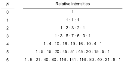
7 1 : 7 : 27 : 61 : 120 : 196 : 257 : 257 : 196 : 120 : 61 : 27 : 7 : 1
8 1 : 8 : 34 : 88 :181 :316 :453 : 514 :453 : 316 : 181 : 88 : 34 : 8 : 1
A can be seen the most intense peak is 514 with respect to the first which is 1 and so in the figure only 15 lines are clearly visible with the first and last lost in the signal to noise of the spectrum.
Add Answer to:
The EPR spectrum of a cyclo-tetrasilane radical anion is shown
below. a) Explain the observed coupling....
2. Below is the observed ESR spectrum of the butadiene anion radical. Interpret the spectrum (i.e.,...
 2. Below is the observed ESR spectrum of the butadiene anion radical. Interpret the spectrum (i.e., What nucleus / nuclei are responsible for coupling with the unpaired electron? Also explain the origins of the multiplet of multiplets, the number of signals in each multiplet, and the relative signal intensities within each multiplet.)
Using the tree approach for predicting first-order NMR spectra for molecules and draw the request...
 Using the tree approach for predicting first-order NMR spectra
for molecules and draw the requested spectra for the following
molecules #1-#3 in the space provided (do not use backside of
pages). All spectra are collected with no {1H} (proton decoupling).
The NMR active nuclei are all I = 1⁄2, i.e., dipolar, so Pascal’s
triangle applies. Consider only 1-bond coupling constants 1JAB, and
ignore ≥2- bond coupling constants (EXCEPT where noted in #2);
label the chosen coupling constant on each branch...
Using the tree approach for predicting first-order NMR spectra
for molecules and draw the requested spectra for the following
molecules #1-#3 in the space provided (do not use backside of
pages). All spectra are collected with no {1H} (proton decoupling).
The NMR active nuclei are all I = 1⁄2, i.e., dipolar, so Pascal’s
triangle applies. Consider only 1-bond coupling constants 1JAB, and
ignore ≥2- bond coupling constants (EXCEPT where noted in #2);
label the chosen coupling constant on each branch...
 2. Below is the observed ESR spectrum of the butadiene anion radical. Interpret the spectrum (i.e., What nucleus / nuclei are responsible for coupling with the unpaired electron? Also explain the origins of the multiplet of multiplets, the number of signals in each multiplet, and the relative signal intensities within each multiplet.)
 Using the tree approach for predicting first-order NMR spectra
for molecules and draw the requested spectra for the following
molecules #1-#3 in the space provided (do not use backside of
pages). All spectra are collected with no {1H} (proton decoupling).
The NMR active nuclei are all I = 1⁄2, i.e., dipolar, so Pascal’s
triangle applies. Consider only 1-bond coupling constants 1JAB, and
ignore ≥2- bond coupling constants (EXCEPT where noted in #2);
label the chosen coupling constant on each branch...
Using the tree approach for predicting first-order NMR spectra
for molecules and draw the requested spectra for the following
molecules #1-#3 in the space provided (do not use backside of
pages). All spectra are collected with no {1H} (proton decoupling).
The NMR active nuclei are all I = 1⁄2, i.e., dipolar, so Pascal’s
triangle applies. Consider only 1-bond coupling constants 1JAB, and
ignore ≥2- bond coupling constants (EXCEPT where noted in #2);
label the chosen coupling constant on each branch...
Most questions answered within 3 hours.
-
1. In a labor market, marginal cost for a firm is
____________.
a. recruiting cost
b....
asked 30 minutes ago -
On January 1, 2019, ABC Company issued $60,000,000 of 20-year,
10.5% bonds when the market rate...
asked 55 minutes ago -
39.4% of US homes continue to use a landline in addition to cell
phone service. 3...
asked 1 hour ago -
Starting with benzene, synthesize 1-phenyl-1-butyne.
Show intermediates and reagents.
asked 2 hours ago -
Create a 32-run crossed array design with six control factors
and two noise factors such that...
asked 3 hours ago -
A 500g sample of sand from source A has the following amounts
retained on each sieve....
asked 3 hours ago -
In
your own words, please explain the essay by John Keynes wrote "The
End of Laissez...
asked 3 hours ago -
How are the matrix and pixels related? Why are smaller
pixels better for diagnostic quality?
asked 3 hours ago -
2. An AC generator has 80 rectangular loops on
its armature. Each loop is 11 cm...
asked 3 hours ago -
Please help me with this question. Consider Aldi’s current and
potential geographic markets (see Exhibit 4...
asked 3 hours ago -
What are the main components of the fermentation process and
give an explanation of each? Include...
asked 3 hours ago -
Explain which types of cells in the body (belonging to which
organs, etc.) are sensitive to...
asked 3 hours ago