
Homework Answers
If you found this answer helpful then please rate it. Comment if you have any problem in understanding this answer.
Add Answer to:
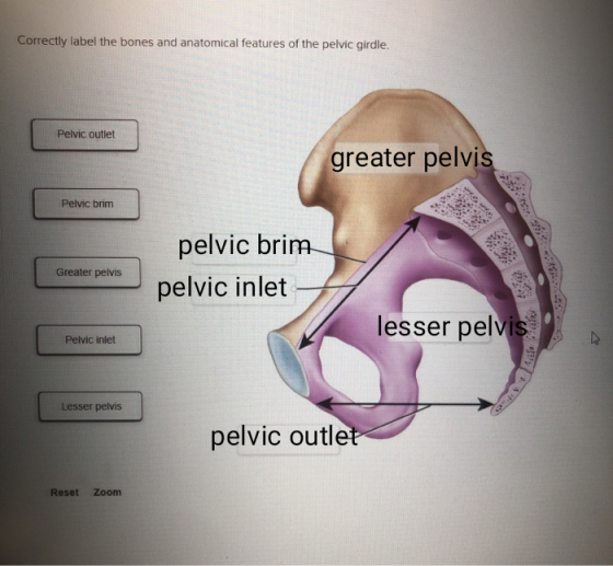
please label
Correctly label the bones and anatomical features of the pelvic girdle. Pelvic outlet Pelvic...
ework Help Correctly label the following anatomical features of the stomach. Cardial part Duodenum Antrum Pylorus...
 ework Help Correctly label the following anatomical features of the stomach. Cardial part Duodenum Antrum Pylorus Gustic russe Pyloric canal Greater omentum Fundic region Pyloric part Pyloric sphincter Reset Zoom < Prev 31 of 50 Neuet >
Label the bones and anatomical features of the hip and thigh - Tufts Medic..Chapter & xQ...
 Label the bones and anatomical features of the hip and thigh
- Tufts Medic..Chapter & xQ Flashcards A. QFlashcands C. QChapter 10 tem Part 2 Homewor.. Help Save&Exit Submit Check my work pones and anatomical features of the hip and thigh. atella occyx Sacrum Tibia Coccyx Fibula Hip bone Lumbar vertebra elvic girdie Femur Tibia Sacrum Reset Zoom < Prev14 of 27 Next> DOLL
Label the bones and anatomical features of the hip and thigh
- Tufts Medic..Chapter & xQ Flashcards A. QFlashcands C. QChapter 10 tem Part 2 Homewor.. Help Save&Exit Submit Check my work pones and anatomical features of the hip and thigh. atella occyx Sacrum Tibia Coccyx Fibula Hip bone Lumbar vertebra elvic girdie Femur Tibia Sacrum Reset Zoom < Prev14 of 27 Next> DOLL
Correctly label the following anatomical features of the coxal joint. Greater trochanter Lesser tubercle Pubofemoral ligament...
 Correctly label the following anatomical features of the coxal joint. Greater trochanter Lesser tubercle Pubofemoral ligament Femur Pubis llium Femoral head Ischium lliofemoral ligament Greater tubercle Lesser trochanter
civity 6 Saved Describe parts of the pelvic girdle by clicking and dragging the labels to...
 civity 6 Saved Describe parts of the pelvic girdle by clicking and dragging the labels to accurately complete each statement pubic symphysis The pelvic girdle consists of two ischial spines The head of the femur articulates with the of the hip bone. is the largest portion of the hip bone. hip bones The distance between the represents the shortest diameter of the pelvic outlet. acetabulum The public bones come together anteriorly to form a cartilaginous joint called the um Reset
work Correctly label the following anatomical features of the oral cavity. Sublingual ortice Upper io Palaopharyngeal...
 work Correctly label the following anatomical features of the oral cavity. Sublingual ortice Upper io Palaopharyngeal Palatino tone Lower lip Lingual trenulum Superofimia! Tongue inferior labial i Il frenulum Submandibular orifice Reset Zoom < Prev 23 of 50 ! Next >
correctly label the following anatomical features of a tibiofemural joint Correctly label the following anatomical features...
 correctly label the following anatomical features of a tibiofemural
joint
Correctly label the following anatomical features of the tibiofemoral joint. Fibular collateral ligament Fibula Patellar ligament (cut) Posterior cruciate ligament Anterior cruciate ligament Tibial collateral ligament Lateral meniscus Medial meniscus Tibia Femur (a) Anterior view Lateral condyle This tissue attaches the patella to the tibia. < Prev 14 of 15 !!! Next >
correctly label the following anatomical features of a tibiofemural
joint
Correctly label the following anatomical features of the tibiofemoral joint. Fibular collateral ligament Fibula Patellar ligament (cut) Posterior cruciate ligament Anterior cruciate ligament Tibial collateral ligament Lateral meniscus Medial meniscus Tibia Femur (a) Anterior view Lateral condyle This tissue attaches the patella to the tibia. < Prev 14 of 15 !!! Next >
Check my work correctly label the following anatomical features or the neart. Hiace your cursor on...
 Check my work correctly label the following anatomical features or the neart. Hiace your cursor on the boxes for more information left pulmonary artery na cava superior vena cava left atrium right coronary artery 2.67 points to pulmonary veins right ventricle eBook pulmonary trunk superior vena cava left ventricle References left ventricle aorta right atrium right ventricle left coronary artery right pulmonary veins right pulmonary artery left atrium inferior vena cava Reset Zoom
Lab 31 The Heart Seved Correctly label the following anatomical features of the heart and thoracic...
 Lab 31 The Heart Seved Correctly label the following anatomical features of the heart and thoracic cage. 13 Right atrium 0.37 points Apex of the heart Skipped Right ventricle Superior vena cava References Pulmonary trunk Reset
Correctly label the following anatomical features of the tibiofemoral joint. Fibula Patellar ligament (cut) Patellar surface...
 Correctly label the following anatomical features of the tibiofemoral joint. Fibula Patellar ligament (cut) Patellar surface Femur Lateral condyle Medial condyle | Lateral meniscus Transverse ligament Tibia (a) Anterior view This is the bone of the upper leg.
Saved omework Assignment Label the bones of the skull in inferior view. Maxilla Palatine bone Zygomatic...
 Saved omework Assignment Label the bones of the skull in inferior view. Maxilla Palatine bone Zygomatic arch Occipital bone Sphenoid bone Greater wing of sphenoid Vomer Temporal bone Parietal bone Reset Zoom
 ework Help Correctly label the following anatomical features of the stomach. Cardial part Duodenum Antrum Pylorus Gustic russe Pyloric canal Greater omentum Fundic region Pyloric part Pyloric sphincter Reset Zoom < Prev 31 of 50 Neuet >
 Label the bones and anatomical features of the hip and thigh
- Tufts Medic..Chapter & xQ Flashcards A. QFlashcands C. QChapter 10 tem Part 2 Homewor.. Help Save&Exit Submit Check my work pones and anatomical features of the hip and thigh. atella occyx Sacrum Tibia Coccyx Fibula Hip bone Lumbar vertebra elvic girdie Femur Tibia Sacrum Reset Zoom < Prev14 of 27 Next> DOLL
Label the bones and anatomical features of the hip and thigh
- Tufts Medic..Chapter & xQ Flashcards A. QFlashcands C. QChapter 10 tem Part 2 Homewor.. Help Save&Exit Submit Check my work pones and anatomical features of the hip and thigh. atella occyx Sacrum Tibia Coccyx Fibula Hip bone Lumbar vertebra elvic girdie Femur Tibia Sacrum Reset Zoom < Prev14 of 27 Next> DOLL
 Correctly label the following anatomical features of the coxal joint. Greater trochanter Lesser tubercle Pubofemoral ligament Femur Pubis llium Femoral head Ischium lliofemoral ligament Greater tubercle Lesser trochanter
 civity 6 Saved Describe parts of the pelvic girdle by clicking and dragging the labels to accurately complete each statement pubic symphysis The pelvic girdle consists of two ischial spines The head of the femur articulates with the of the hip bone. is the largest portion of the hip bone. hip bones The distance between the represents the shortest diameter of the pelvic outlet. acetabulum The public bones come together anteriorly to form a cartilaginous joint called the um Reset
 work Correctly label the following anatomical features of the oral cavity. Sublingual ortice Upper io Palaopharyngeal Palatino tone Lower lip Lingual trenulum Superofimia! Tongue inferior labial i Il frenulum Submandibular orifice Reset Zoom < Prev 23 of 50 ! Next >
 correctly label the following anatomical features of a tibiofemural
joint
Correctly label the following anatomical features of the tibiofemoral joint. Fibular collateral ligament Fibula Patellar ligament (cut) Posterior cruciate ligament Anterior cruciate ligament Tibial collateral ligament Lateral meniscus Medial meniscus Tibia Femur (a) Anterior view Lateral condyle This tissue attaches the patella to the tibia. < Prev 14 of 15 !!! Next >
correctly label the following anatomical features of a tibiofemural
joint
Correctly label the following anatomical features of the tibiofemoral joint. Fibular collateral ligament Fibula Patellar ligament (cut) Posterior cruciate ligament Anterior cruciate ligament Tibial collateral ligament Lateral meniscus Medial meniscus Tibia Femur (a) Anterior view Lateral condyle This tissue attaches the patella to the tibia. < Prev 14 of 15 !!! Next >
 Check my work correctly label the following anatomical features or the neart. Hiace your cursor on the boxes for more information left pulmonary artery na cava superior vena cava left atrium right coronary artery 2.67 points to pulmonary veins right ventricle eBook pulmonary trunk superior vena cava left ventricle References left ventricle aorta right atrium right ventricle left coronary artery right pulmonary veins right pulmonary artery left atrium inferior vena cava Reset Zoom
 Lab 31 The Heart Seved Correctly label the following anatomical features of the heart and thoracic cage. 13 Right atrium 0.37 points Apex of the heart Skipped Right ventricle Superior vena cava References Pulmonary trunk Reset
 Correctly label the following anatomical features of the tibiofemoral joint. Fibula Patellar ligament (cut) Patellar surface Femur Lateral condyle Medial condyle | Lateral meniscus Transverse ligament Tibia (a) Anterior view This is the bone of the upper leg.
 Saved omework Assignment Label the bones of the skull in inferior view. Maxilla Palatine bone Zygomatic arch Occipital bone Sphenoid bone Greater wing of sphenoid Vomer Temporal bone Parietal bone Reset Zoom
Most questions answered within 3 hours.
-
Calculate the number density of argon gas at a temperature of
24C and a pressure of...
asked 2 hours ago -
Alternative
Classification
How to Estimate
Probabilities from Data? ( For continuous Attributes)
And How to generate...
asked 2 hours ago -
An explosion breaks a 20.0-kg object into three parts. The
object is initially moving at a...
asked 3 hours ago -
Calculate the approximate number of residues of Rubisco, which
is involved in carbon fixation in plants,...
asked 4 hours ago -
Other decisions about scientific claims can have a much broader
impact.ENERGYarrow-10x10.png, environment, health, security - all...
asked 4 hours ago -
I need to write a research paper and work cited about this
topic: The United States...
asked 5 hours ago -
Hello! I was wondering if I could have some help?
If the vapor pressure of carvone...
asked 5 hours ago -
An economist wants to estimate the mean per capita income (in
thousands of dollars) for a...
asked 6 hours ago -
What would be the input/output characteristic of a circuit
obtained by putting two of your 2's-complementers...
asked 6 hours ago -
In Drosophila, the transition from the syncytial blastoderm
stage to the cellular blastoderm stage is a...
asked 6 hours ago -
Project management question:
Name 3 different types of resources (hint: humans are one
type)
asked 6 hours ago -
Consider the following reaction: C 2H 2( g) + 2H 2( g) C 2H 6(
g)...
asked 6 hours ago