Pre-Lab #4 (This will help you prepare for the Skeletal Anatomy Test at end of Week...
Pre-Lab #4 (This will help you prepare for the Skeletal Anatomy Test at end of Week 5).
-
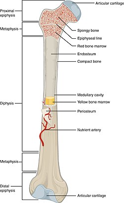
Diagram (or obtain an unlabeled digital image of) and label the bones below. You can obtain unlabeled images online.
- For each bone complete the following tasks for Pre-Lab #4
- Identify the bone and whether it is right or left (if that applies).
- Identify any markings or structures listed below on the bone (ex: fossa, cavity, process, etc).
- Identify any specific joints/articulations this bone is found in and other bones involved in the articulation.
- Explain how to differentiate between a male and female pelvis.
- For each bone complete the following tasks for Pre-Lab #4
Appendicular - Shoulder Region
Clavicle
Scapula
- spine of the scapula
- medial, lateral, and superior borders
- superior angle
- inferior angle
- acromion (acromial process)
- coracoid process
- supraspinous fossa
- infraspinous fossa
- subscapular fossa
- glenoid cavity (glenoid fossa)
Humerus
- head of the humerus
- greater tubercle
- lesser tubercle
- anatomical neck
- surgical neck
- deltoid tuberosity
- trochlea
- capitulum
- olecranon fossa
- medial epicondyle
- lateral epicondyle
Arms to Hand
Ulna
- styloid process of the ulna
- olecranon
- trochlear (semilunar) notch
Radius
- styloid process of the radius
Carpals
- (on an articulated hand only; you need to identify each one)
- Scaphoid, lunate, pisiform, hamate, triquetrum, trapezoid, capitate, trapezium
Metacarpals
Phalanges
Pelvic Girdle
Coxal bone(s)
- os(sa) coxa(e)
- ilium
- ischium
- pubis
- acetabulum
- obturator foramen
ilium
- iliac spine
- iliac crest
- iliac fossa (on the anterior surface only)
- greater sciatic notch
ischium
- ischial (sciatic) spine
- ischial ramus
- lesser sciatic notch
pubis
- superior ramus of the pubis
- inferior ramus of the pubis
- pubic symphyseal surface
- Pubic symphysis
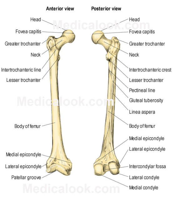
femur
- head of the femur
- neck of the femur
- greater trochanter
- lesser trochanter
- medial condyle
- lateral condyle
- medial epicondyle
- lateral epicondyle
- gluteal tuberosity
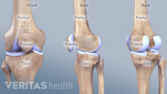
patella
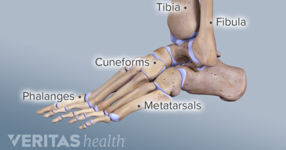
tibia
- medial condyle
- lateral condyle
- tibial tuberosity
- medial malleolus
fibula
- head of the fibula
- lateral malleolus
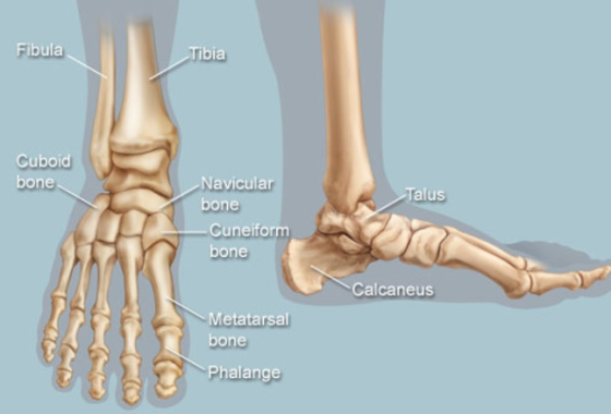
tarsals
- (on an articulated foot; you need to identify each one)
- calcaneus, talus, cuneiforms (medial, middle and lateral), cuboid, navicular
metatarsals
phalanges
Homework Answers
Human skeleton made up of 206 bones.It functions as a support system of the body, gives the shape of the body, provides protection to organs and other systems of the body, to provide attachments for muscles, to produce movement and to produce Red blood cells.
The pelvis is one of the most useful skeletal element for differentiating between male and female. Female pelves are larger and wider than male pelves and have round pelvic inlet. The male iliac crest is higher than female, causing their false pelves to look taller and narrower.
Below are labeled the diagram.


Add Answer to:
Pre-Lab #4 (This will help you prepare for the Skeletal Anatomy
Test at end of Week...
Drag the appropriate labels to their respective targets. Reset Help Obturator foramen lliac crest Pubis Anterior...
 Drag the appropriate labels to their respective targets. Reset Help Obturator foramen lliac crest Pubis Anterior inferior iliac spine Anterior superior iliac spine Ischium Posterior superior iliac spine Greater sciatic notch Posterior inferior iliac spine Acetabulum mium llium Lesser sciatic notch Ischial tuberosity Ischial tuberosity Right pelvic bone, medial view Right pelvic bone, lateral view
label the parts on the humerus: head,greater tubercle 4. Label the parts on the humerus: head,...
 label the parts on the humerus: head,greater tubercle
4. Label the parts on the humerus: head, greater tubercle, lesser tubercle, intertubercular sulcus, deltoid tuberosity, olecranon fossa, coronoid fossa, trochlea, capitulum, medial epicondyle, lateral epicondyle head ofd humerus head of humeus greater tubercle lesser tubercle -deltoid tuberosity deltold tuberosity coronoid fossa olecranon fossa lateral epicondyle medial epycondyle medial -lateral epvcondyle trochlea trochlea Anterior view Posterior view 5. Label the two bones located in the forearm and identify specific parts of their...
label the parts on the humerus: head,greater tubercle
4. Label the parts on the humerus: head, greater tubercle, lesser tubercle, intertubercular sulcus, deltoid tuberosity, olecranon fossa, coronoid fossa, trochlea, capitulum, medial epicondyle, lateral epicondyle head ofd humerus head of humeus greater tubercle lesser tubercle -deltoid tuberosity deltold tuberosity coronoid fossa olecranon fossa lateral epicondyle medial epycondyle medial -lateral epvcondyle trochlea trochlea Anterior view Posterior view 5. Label the two bones located in the forearm and identify specific parts of their...
please answer the question as in the picture(by filling the table and note in paragraphs or sentences) NAME: FUNCTIO...
 please answer the question as in the picture(by
filling the table and note in paragraphs or sentences)
NAME: FUNCTIONS OF BONE MARKINGS MARKING BONE(S) MARKING FUNCTION articulates with it, what OCCURS ON (e.g s through it or attaches to it) External auditory meatus Mastoid process Styloid process Petrous portion Internal auditory meatus Foramen magnum Occipital condyle Sella turcica Op plate Crista galli cess Intervertebral discs Zygomatic process Temporal process Condylar process Mandibular fossa Superior articular processes Inferior articular processes Transverse...
please answer the question as in the picture(by
filling the table and note in paragraphs or sentences)
NAME: FUNCTIONS OF BONE MARKINGS MARKING BONE(S) MARKING FUNCTION articulates with it, what OCCURS ON (e.g s through it or attaches to it) External auditory meatus Mastoid process Styloid process Petrous portion Internal auditory meatus Foramen magnum Occipital condyle Sella turcica Op plate Crista galli cess Intervertebral discs Zygomatic process Temporal process Condylar process Mandibular fossa Superior articular processes Inferior articular processes Transverse...
please help Humerus Label the following: Head (2x) Anatomical neck (2x) Surgical neck Greater tubercle (2x)...
 please help
Humerus Label the following: Head (2x) Anatomical neck (2x) Surgical neck Greater tubercle (2x) Lesser tubercle Intertubercular sulcus Deltoid tuberosity Radial fossa Capitulum Coronoid fossa Medial epicondyle (2x) Lateral epicondyle Trochlea (2x) Olecranon fossa -Head - Anatomical neck Head- Anatomical- neck Greater tubercle -Surgical neuk -Delted القاروطي Coronoid fossa Radial fossa -Medial eplondyle Torochlea Capttulum 0L_OFor) fossa Medial epicondyle - Lateral Trochlea opitonduk
please help
Humerus Label the following: Head (2x) Anatomical neck (2x) Surgical neck Greater tubercle (2x) Lesser tubercle Intertubercular sulcus Deltoid tuberosity Radial fossa Capitulum Coronoid fossa Medial epicondyle (2x) Lateral epicondyle Trochlea (2x) Olecranon fossa -Head - Anatomical neck Head- Anatomical- neck Greater tubercle -Surgical neuk -Delted القاروطي Coronoid fossa Radial fossa -Medial eplondyle Torochlea Capttulum 0L_OFor) fossa Medial epicondyle - Lateral Trochlea opitonduk
ACTIVITY 2 Upper Limb: Ex amining the Bones of the Arm and Forearm The humerus is...
 ACTIVITY 2 Upper Limb: Ex amining the Bones of the Arm and Forearm The humerus is the only bone found in the arm. It is the longest and largest bone of the upper limb. O On the skeleton, locate the humerus in each arm. Notice that it is involved in the formation of two major joints: the shoulder joint and the elbow joint Posterior Anterior 2 On a skeleton or disarticulated humerus, identify the bone markings Greater labeled in the...
Art-labeling Activity: Bones of the ankle and foot (superior view, right foot) Reset Help Lateral cuneiform...
 Art-labeling Activity: Bones of the ankle and foot (superior view, right foot) Reset Help Lateral cuneiform Calcaneus Distal phalan Cuboid Talus Metatarsal bones I HII Proximal phalanx Intermediate cuneiform Medial cuneiform Middle phalanx Navicular Art-labeling Activity: The Major Bones of the Appendicular Skeleton (lower limb) llum Phalanges Tarsal bones Metatarsals Femur Tibia Pubis Ischium Patola Fibula Art-labeling Activity: Bone Markings (Femur) Reset Help Head Neck Trochanter Tubercle Condyle Facet
The r Skeleton TIONS 34-39: Identify the following bone markings by palpating them on yoursel lowing...
 The r Skeleton TIONS 34-39: Identify the following bone markings by palpating them on yoursel lowing list. Answers may be used once or not at all. f or your lab partner. Select your answers from a. patella b. greater trochanter c. lateral malleolus d. iliac crest e. medial condyle f. tibial tuberosity g. lesser trochanter h. ischial tuberosity i. lateral condyle j. medial malleolus k. patellar surface I. head of the fibula 34. Press gently with your fingers along your...
 Drag the appropriate labels to their respective targets. Reset Help Obturator foramen lliac crest Pubis Anterior inferior iliac spine Anterior superior iliac spine Ischium Posterior superior iliac spine Greater sciatic notch Posterior inferior iliac spine Acetabulum mium llium Lesser sciatic notch Ischial tuberosity Ischial tuberosity Right pelvic bone, medial view Right pelvic bone, lateral view
 label the parts on the humerus: head,greater tubercle
4. Label the parts on the humerus: head, greater tubercle, lesser tubercle, intertubercular sulcus, deltoid tuberosity, olecranon fossa, coronoid fossa, trochlea, capitulum, medial epicondyle, lateral epicondyle head ofd humerus head of humeus greater tubercle lesser tubercle -deltoid tuberosity deltold tuberosity coronoid fossa olecranon fossa lateral epicondyle medial epycondyle medial -lateral epvcondyle trochlea trochlea Anterior view Posterior view 5. Label the two bones located in the forearm and identify specific parts of their...
label the parts on the humerus: head,greater tubercle
4. Label the parts on the humerus: head, greater tubercle, lesser tubercle, intertubercular sulcus, deltoid tuberosity, olecranon fossa, coronoid fossa, trochlea, capitulum, medial epicondyle, lateral epicondyle head ofd humerus head of humeus greater tubercle lesser tubercle -deltoid tuberosity deltold tuberosity coronoid fossa olecranon fossa lateral epicondyle medial epycondyle medial -lateral epvcondyle trochlea trochlea Anterior view Posterior view 5. Label the two bones located in the forearm and identify specific parts of their...
 please answer the question as in the picture(by
filling the table and note in paragraphs or sentences)
NAME: FUNCTIONS OF BONE MARKINGS MARKING BONE(S) MARKING FUNCTION articulates with it, what OCCURS ON (e.g s through it or attaches to it) External auditory meatus Mastoid process Styloid process Petrous portion Internal auditory meatus Foramen magnum Occipital condyle Sella turcica Op plate Crista galli cess Intervertebral discs Zygomatic process Temporal process Condylar process Mandibular fossa Superior articular processes Inferior articular processes Transverse...
please answer the question as in the picture(by
filling the table and note in paragraphs or sentences)
NAME: FUNCTIONS OF BONE MARKINGS MARKING BONE(S) MARKING FUNCTION articulates with it, what OCCURS ON (e.g s through it or attaches to it) External auditory meatus Mastoid process Styloid process Petrous portion Internal auditory meatus Foramen magnum Occipital condyle Sella turcica Op plate Crista galli cess Intervertebral discs Zygomatic process Temporal process Condylar process Mandibular fossa Superior articular processes Inferior articular processes Transverse...
 please help
Humerus Label the following: Head (2x) Anatomical neck (2x) Surgical neck Greater tubercle (2x) Lesser tubercle Intertubercular sulcus Deltoid tuberosity Radial fossa Capitulum Coronoid fossa Medial epicondyle (2x) Lateral epicondyle Trochlea (2x) Olecranon fossa -Head - Anatomical neck Head- Anatomical- neck Greater tubercle -Surgical neuk -Delted القاروطي Coronoid fossa Radial fossa -Medial eplondyle Torochlea Capttulum 0L_OFor) fossa Medial epicondyle - Lateral Trochlea opitonduk
please help
Humerus Label the following: Head (2x) Anatomical neck (2x) Surgical neck Greater tubercle (2x) Lesser tubercle Intertubercular sulcus Deltoid tuberosity Radial fossa Capitulum Coronoid fossa Medial epicondyle (2x) Lateral epicondyle Trochlea (2x) Olecranon fossa -Head - Anatomical neck Head- Anatomical- neck Greater tubercle -Surgical neuk -Delted القاروطي Coronoid fossa Radial fossa -Medial eplondyle Torochlea Capttulum 0L_OFor) fossa Medial epicondyle - Lateral Trochlea opitonduk
 ACTIVITY 2 Upper Limb: Ex amining the Bones of the Arm and Forearm The humerus is the only bone found in the arm. It is the longest and largest bone of the upper limb. O On the skeleton, locate the humerus in each arm. Notice that it is involved in the formation of two major joints: the shoulder joint and the elbow joint Posterior Anterior 2 On a skeleton or disarticulated humerus, identify the bone markings Greater labeled in the...
 Art-labeling Activity: Bones of the ankle and foot (superior view, right foot) Reset Help Lateral cuneiform Calcaneus Distal phalan Cuboid Talus Metatarsal bones I HII Proximal phalanx Intermediate cuneiform Medial cuneiform Middle phalanx Navicular Art-labeling Activity: The Major Bones of the Appendicular Skeleton (lower limb) llum Phalanges Tarsal bones Metatarsals Femur Tibia Pubis Ischium Patola Fibula Art-labeling Activity: Bone Markings (Femur) Reset Help Head Neck Trochanter Tubercle Condyle Facet
 The r Skeleton TIONS 34-39: Identify the following bone markings by palpating them on yoursel lowing list. Answers may be used once or not at all. f or your lab partner. Select your answers from a. patella b. greater trochanter c. lateral malleolus d. iliac crest e. medial condyle f. tibial tuberosity g. lesser trochanter h. ischial tuberosity i. lateral condyle j. medial malleolus k. patellar surface I. head of the fibula 34. Press gently with your fingers along your...
Most questions answered within 3 hours.
-
3) What are the typical social structures in a global city?
asked 17 minutes ago -
Luther Corporation
Consolidated Balance Sheet
December 31, 2019 and 2018 (in $ millions)
Assets
2019
2018...
asked 19 minutes ago -
(Expected rate of return and risk) Carter Inc. is evaluating a
security. Calculate the investment’s expected...
asked 3 hours ago -
What specific indicators can point to lack of progress for
African Americans in American society?
asked 4 hours ago -
1-The Electrons in a beam are moving at 2.7×10⁸ m/s in an
electric field of 15000...
asked 4 hours ago -
A gas tank is a vertical cylinder. It has a radius of 1m, a
height of...
asked 4 hours ago -
Accent Software faces the following conditions. All of these
support Accent’s use of a market-penetration pricing...
asked 5 hours ago -
A mathematically inclined friend emails you the following
instructions: "Meet me in the cafeteria the first...
asked 5 hours ago -
A monopoly sells in two countries . The demand curves in the two
countries are p1...
asked 6 hours ago -
A .15kg rubber ball is bounced off a wall. Before hitting the
wall, the ball moves...
asked 7 hours ago -
A manufacturing company preparing to build a new plant is
considering three potential locations for it....
asked 7 hours ago -
B. If compound Y has approximately the same values of solubility
in toluene as compound X,...
asked 8 hours ago