

please I need your help with this question. thanks
Styles Paragraph Editing This image is from the dermis of the skin. Remember from Ch 1 that the integument has a number of important homeostatic functions. These blood vessels which are found in the dermis transport gases and nutrients, but they are also well adapted to This requires the ability of the arteries and arterioles to both withstand the high systolic blood pressure and to control the rate of blood flow through the tissues. They accomplish this by having thicker layers of elastic connective tissue and smooth muscle fibers. Letters A and B indicate () while F is a ( ). Examine the contents of letters A, B, and F carefully. They are all ). Examine the thin wall of vessel A. It has ) layers. The letter D arrows are pointing toward nuclei of L). The main tissue which comprises the dermis is ( ).The reddish-violet structures which make up the majority of the tissue are() .They were produced by cells that manufactured the monomers (building blocks) and enzymes that joined the monomers together (polymerization) outside of the cell. The cells are known as Their purple nuclei are visible scattered though the dermis
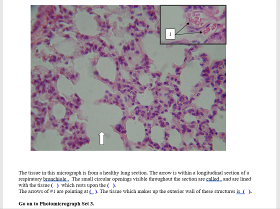
1 The tissue in this micrograph is from a healthy lung section. The arrow is within a longitudinal section of a respiratory bronchiole. The small circular openings visible throughout the section are called, and are lined with the tissue ( ) which rests upon the ( ). The arrows of #1 are pointing at (). The tissue which makes up the exterior wall of these structures is (). Go on to Photomicrograph Set 3.
Homework Answers
Ans. The dermis is the middle layer of the skin, between epidermis and subcutaneous layer. This layer is richly supplied with blood vessels which carry out the function of exchange of gases and nutrients. Along with this they are also adapted to stress and strain. So, in case of first blank, we can write stress as the answer.
Letters A and B indicate capillaries while letter F indicates a vein. When we examine the thin wall of vessel A, it has capillary endothelial layers.
The letter D arrows arec pointing towards the nuclei of endothelial cells.
The main tissue which comprises the dermis is connective tissue. The reddish violet structures that make up the majority of the tissues are collagens. The cells that make collagens are called fibroblasts.
Ans. The small circular openings visible through out the section are alveoli and are lined with the tissue alveolar epithelium, resting upon the basement membrane.
The arrows of #1 are pointing at capillaries. The tissue which makes up the exterior wall of this structures is capillary endothelium.
Add Answer to:
please I need your help with this question. thanks Photomicrograph Set 2.An excellent reference for this...
please i need your help with this question. thanks The micrographs in this document are from...
 please i need your help with this question. thanks
The micrographs in this document are from a cross-section of the small intestine. This image is part of the field of view using a 10X eyepiece and a 4X objective, providing a total magnification of 40X. The digital camera image here is approximately 2/3 of the visual field of view, giving an actual magnification of about 60X. What tissues are visible here? For a closer look, change the size of the...
please i need your help with this question. thanks
The micrographs in this document are from a cross-section of the small intestine. This image is part of the field of view using a 10X eyepiece and a 4X objective, providing a total magnification of 40X. The digital camera image here is approximately 2/3 of the visual field of view, giving an actual magnification of about 60X. What tissues are visible here? For a closer look, change the size of the...
can you please answer all, i need to study this. please and thank you 2. Can...
 can you please answer all, i need to study this. please and
thank you
2. Can you think of a way to remember the relative locations of the three layers2) a. () b. 3. Explain why the tunica media is so thicker in the artery g much than in the vein. C d 4. How do the structures of the blood vessels the largest change arteries divide, becoming smaller arteries, then arterioles capillaries, then ever larger veins. as 5. How...
can you please answer all, i need to study this. please and
thank you
2. Can you think of a way to remember the relative locations of the three layers2) a. () b. 3. Explain why the tunica media is so thicker in the artery g much than in the vein. C d 4. How do the structures of the blood vessels the largest change arteries divide, becoming smaller arteries, then arterioles capillaries, then ever larger veins. as 5. How...
I need new and unique answers, please. (Use your own words, don't copy and paste), Please...
I need new and unique answers, please. (Use your own words, don't copy and paste), Please Use your keyboard (Don't use handwriting) Thank you.. BIOL 102 Choose any system of the human body and prepare a response to the following questions in 1-2 pages: 1. Introduction (Explain the system with the components) 2. Body (Explain how the system relates to achieve homeostasis in human body) 3. Conclusion (Choose any disease common in Kingdom of Saudi Arabia and explain how and...
I need your help ASAP! please show all work for this activity sheet f True/False Write...
 I
need your help ASAP! please show all work for this activity sheet
f True/False Write T for true, F for false a) Prostaglandins are used to make steroid hormones b) Prostaglandins are synthesized only in the liver c) Prostaglandins can be used to induce labor d) Prostaglandins are synthesized in the cell only when it is needed e) Prostaglandins prevent the inflammation response Prostaglandins can cause fever and pain sensitivity g) Phospholipids are the only lipids found in a...
I
need your help ASAP! please show all work for this activity sheet
f True/False Write T for true, F for false a) Prostaglandins are used to make steroid hormones b) Prostaglandins are synthesized only in the liver c) Prostaglandins can be used to induce labor d) Prostaglandins are synthesized in the cell only when it is needed e) Prostaglandins prevent the inflammation response Prostaglandins can cause fever and pain sensitivity g) Phospholipids are the only lipids found in a...
Can you please help me to find Possible test questions? Course Here.com Test #4 " Autonomic...
 Can you please help me to find Possible test questions?
Course Here.com Test #4 " Autonomic Nervous System Overview of the Autonomic Nervous System (ANSH Maior Functions: maintain optimal muscle in order to maintain homeostatic state within the body Aalso is inv performance of visceral organs, glands, smooth muscle, and cardiac not under conscious control: regulates heart rate, blood pressure, MOST "effectors" (organs & tissues regulated) are visceral- r function, and secretions emperaturs smooth musele contraction, glandula most are not...
Can you please help me to find Possible test questions?
Course Here.com Test #4 " Autonomic Nervous System Overview of the Autonomic Nervous System (ANSH Maior Functions: maintain optimal muscle in order to maintain homeostatic state within the body Aalso is inv performance of visceral organs, glands, smooth muscle, and cardiac not under conscious control: regulates heart rate, blood pressure, MOST "effectors" (organs & tissues regulated) are visceral- r function, and secretions emperaturs smooth musele contraction, glandula most are not...
i really need help with the graphs Driving Can Be Dangerous to Your Health: An Interrupted...
 i really need help with the graphs
Driving Can Be Dangerous to Your Health: An Interrupted Case Study in Physiology Phil Stephens Department of Biology Villanova University Part 1-The Grandparents Arrive Dave pulled the cell phone out of his pocket, cursing himself for not putting it on vibrate. The children, Jason and Laura, were both asleep, and he knew that the rest of the day would not be fun if they were awakened from their naps. "Hi, Dave. We're just...
i really need help with the graphs
Driving Can Be Dangerous to Your Health: An Interrupted Case Study in Physiology Phil Stephens Department of Biology Villanova University Part 1-The Grandparents Arrive Dave pulled the cell phone out of his pocket, cursing himself for not putting it on vibrate. The children, Jason and Laura, were both asleep, and he knew that the rest of the day would not be fun if they were awakened from their naps. "Hi, Dave. We're just...
 please i need your help with this question. thanks
The micrographs in this document are from a cross-section of the small intestine. This image is part of the field of view using a 10X eyepiece and a 4X objective, providing a total magnification of 40X. The digital camera image here is approximately 2/3 of the visual field of view, giving an actual magnification of about 60X. What tissues are visible here? For a closer look, change the size of the...
please i need your help with this question. thanks
The micrographs in this document are from a cross-section of the small intestine. This image is part of the field of view using a 10X eyepiece and a 4X objective, providing a total magnification of 40X. The digital camera image here is approximately 2/3 of the visual field of view, giving an actual magnification of about 60X. What tissues are visible here? For a closer look, change the size of the...
 can you please answer all, i need to study this. please and
thank you
2. Can you think of a way to remember the relative locations of the three layers2) a. () b. 3. Explain why the tunica media is so thicker in the artery g much than in the vein. C d 4. How do the structures of the blood vessels the largest change arteries divide, becoming smaller arteries, then arterioles capillaries, then ever larger veins. as 5. How...
can you please answer all, i need to study this. please and
thank you
2. Can you think of a way to remember the relative locations of the three layers2) a. () b. 3. Explain why the tunica media is so thicker in the artery g much than in the vein. C d 4. How do the structures of the blood vessels the largest change arteries divide, becoming smaller arteries, then arterioles capillaries, then ever larger veins. as 5. How...
 I
need your help ASAP! please show all work for this activity sheet
f True/False Write T for true, F for false a) Prostaglandins are used to make steroid hormones b) Prostaglandins are synthesized only in the liver c) Prostaglandins can be used to induce labor d) Prostaglandins are synthesized in the cell only when it is needed e) Prostaglandins prevent the inflammation response Prostaglandins can cause fever and pain sensitivity g) Phospholipids are the only lipids found in a...
I
need your help ASAP! please show all work for this activity sheet
f True/False Write T for true, F for false a) Prostaglandins are used to make steroid hormones b) Prostaglandins are synthesized only in the liver c) Prostaglandins can be used to induce labor d) Prostaglandins are synthesized in the cell only when it is needed e) Prostaglandins prevent the inflammation response Prostaglandins can cause fever and pain sensitivity g) Phospholipids are the only lipids found in a...
 Can you please help me to find Possible test questions?
Course Here.com Test #4 " Autonomic Nervous System Overview of the Autonomic Nervous System (ANSH Maior Functions: maintain optimal muscle in order to maintain homeostatic state within the body Aalso is inv performance of visceral organs, glands, smooth muscle, and cardiac not under conscious control: regulates heart rate, blood pressure, MOST "effectors" (organs & tissues regulated) are visceral- r function, and secretions emperaturs smooth musele contraction, glandula most are not...
Can you please help me to find Possible test questions?
Course Here.com Test #4 " Autonomic Nervous System Overview of the Autonomic Nervous System (ANSH Maior Functions: maintain optimal muscle in order to maintain homeostatic state within the body Aalso is inv performance of visceral organs, glands, smooth muscle, and cardiac not under conscious control: regulates heart rate, blood pressure, MOST "effectors" (organs & tissues regulated) are visceral- r function, and secretions emperaturs smooth musele contraction, glandula most are not...
 i really need help with the graphs
Driving Can Be Dangerous to Your Health: An Interrupted Case Study in Physiology Phil Stephens Department of Biology Villanova University Part 1-The Grandparents Arrive Dave pulled the cell phone out of his pocket, cursing himself for not putting it on vibrate. The children, Jason and Laura, were both asleep, and he knew that the rest of the day would not be fun if they were awakened from their naps. "Hi, Dave. We're just...
i really need help with the graphs
Driving Can Be Dangerous to Your Health: An Interrupted Case Study in Physiology Phil Stephens Department of Biology Villanova University Part 1-The Grandparents Arrive Dave pulled the cell phone out of his pocket, cursing himself for not putting it on vibrate. The children, Jason and Laura, were both asleep, and he knew that the rest of the day would not be fun if they were awakened from their naps. "Hi, Dave. We're just...
Most questions answered within 3 hours.
-
3) What are the typical social structures in a global city?
asked 29 minutes ago -
Luther Corporation
Consolidated Balance Sheet
December 31, 2019 and 2018 (in $ millions)
Assets
2019
2018...
asked 31 minutes ago -
(Expected rate of return and risk) Carter Inc. is evaluating a
security. Calculate the investment’s expected...
asked 3 hours ago -
What specific indicators can point to lack of progress for
African Americans in American society?
asked 4 hours ago -
1-The Electrons in a beam are moving at 2.7×10⁸ m/s in an
electric field of 15000...
asked 4 hours ago -
A gas tank is a vertical cylinder. It has a radius of 1m, a
height of...
asked 4 hours ago -
Accent Software faces the following conditions. All of these
support Accent’s use of a market-penetration pricing...
asked 5 hours ago -
A mathematically inclined friend emails you the following
instructions: "Meet me in the cafeteria the first...
asked 5 hours ago -
A monopoly sells in two countries . The demand curves in the two
countries are p1...
asked 6 hours ago -
A .15kg rubber ball is bounced off a wall. Before hitting the
wall, the ball moves...
asked 7 hours ago -
A manufacturing company preparing to build a new plant is
considering three potential locations for it....
asked 7 hours ago -
B. If compound Y has approximately the same values of solubility
in toluene as compound X,...
asked 8 hours ago