How to determine which bacteria was growing on this Mitis salivarius agar plate? Streptococcus salicarius, streptococcus...
How to determine which bacteria was growing on this Mitis salivarius agar plate? Streptococcus salicarius, streptococcus mitis, streptococcus mutans, or enterococcus.
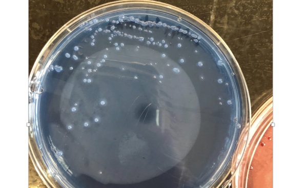
Homework Answers
Strep salivarius is sticky, mucoid, gum-drop
colonies.
Strep mitis colonies are small, flat, light blue.
Strep mutans are irregular shaped with granular
frosted-glass appearance.
Enterococcus are dark, blue-black colonies.
Therfore in the picture it is Streptococcus salivarius.
Add Answer to:
How to determine which bacteria was growing on this Mitis
salivarius agar plate? Streptococcus salicarius, streptococcus...
a catalase test is usually not performed on bacteria growing on a blood agar plate. Why...
a catalase test is usually not performed on bacteria growing on a blood agar plate. Why not?
1)An eosin methylene blue (EMB) agar plate was inoculated with four different species of bacteria. Below...
1)An eosin methylene blue (EMB) agar plate was inoculated with four different species of bacteria. Below are the colony growth after inoculation. Which shows a vigorous lactose fermenter? A) A brown media with an oblong culture of bacteria. The colony shows vigorous growth and is the same color as the media. B) A brown media with an oblong colony of bacteria that experiences weak growth. The color of the media is visible through the translucent white color of the bacterial...
See Hint Consider the colonies growing on this sorbitol MacConkey agar plate. Which component of the...
 See Hint Consider the colonies growing on this sorbitol MacConkey agar plate. Which component of the medium is being metabolized by the ones that appear colorless? Troy Biological BD Worldwide Choose one: A. peptone B. agar C. neutral red D. sorbitol E. sodium chloride < 18/18
What are the advantages of growing bacteria on a plate versus broth? How can you tell...
What are the advantages of growing bacteria on a plate versus broth? How can you tell whether the colonies on a plate are from the original culture and not contaminants?
4) Which of the bacteria grew well in 6.5% NaCl? Which bacteria did not grow well...
4) Which of the bacteria grew well in 6.5% NaCl? Which bacteria did not grow well in 6.5% NaCl? Based on your knowledge of the normal microflora, do these results make sense? Why or why not? The bacterias that grew well in 6.5% NaCl were the Staph bacteria (Staphylococcus aureus, Staphylococcus epidermidis, and Staphylococcus saprophyticus), Micrococcus luteus, and Enterococcus faebalis. The bacterias that did not grow well in 6.5% NaCl were Streptococcus pyogenes and Streptococcus pneumoniae.
How would you verify that the colonies that grew on a MacConkey agar plate were Gram...
How would you verify that the colonies that grew on a MacConkey agar plate were Gram negative? What types of bacteria are inhibited on MacConkey agar?
Please answer all (1 pt 27) The two bacteria cultured on this mannitol salt agar plate...
 Please answer all
(1 pt 27) The two bacteria cultured on this mannitol salt agar plate will have the same reaction. One reacted nearly 24 hours before the other. What substance in the agar was catabolized by both bacteria that produced the demonstrated colorimetric change? Image gallery (click for full size) (1pt 28) The presence of bacterial growth on this mannitol salt agar plate suggests that these bacteria can tolerate the hyperosmotic condition produced by the salt. The term used...
Please answer all
(1 pt 27) The two bacteria cultured on this mannitol salt agar plate will have the same reaction. One reacted nearly 24 hours before the other. What substance in the agar was catabolized by both bacteria that produced the demonstrated colorimetric change? Image gallery (click for full size) (1pt 28) The presence of bacterial growth on this mannitol salt agar plate suggests that these bacteria can tolerate the hyperosmotic condition produced by the salt. The term used...
1. What are 4 standard terms used to describe morphological characteristics of colonies growing on an...
1. What are 4 standard terms used to describe morphological characteristics of colonies growing on an agar plate? 2. Describe the T-streak method 3. Describe which inoculation tool is the most approproiate to use when transfering bacteria from an agar plate to a stab tube. 4. Why is a buffer added to agar? Explain your answer. 5. Look up a growth curve for bacteria growing on an agar plate. Describe why the lag phase occurs.
Growing on EMB Agar 10. EMB agar is (choose as many as apply): – A. differential...
 Growing on EMB Agar 10. EMB agar is (choose as many as apply): – A. differential - B. Selective C. enriched • 11. Identify by genus and species the organisms on the right hand plate in the photo Enterobacter aerogenes
1.) How could MacConkey agar aid in the identification of coliforms? 2.) Streptococcus Uniate ferments mannitol....
1.) How could MacConkey agar aid in the identification of coliforms? 2.) Streptococcus Uniate ferments mannitol. Could MacConkey agar be used to isolate S. iniae from a complex mixture?
 See Hint Consider the colonies growing on this sorbitol MacConkey agar plate. Which component of the medium is being metabolized by the ones that appear colorless? Troy Biological BD Worldwide Choose one: A. peptone B. agar C. neutral red D. sorbitol E. sodium chloride < 18/18
 Please answer all
(1 pt 27) The two bacteria cultured on this mannitol salt agar plate will have the same reaction. One reacted nearly 24 hours before the other. What substance in the agar was catabolized by both bacteria that produced the demonstrated colorimetric change? Image gallery (click for full size) (1pt 28) The presence of bacterial growth on this mannitol salt agar plate suggests that these bacteria can tolerate the hyperosmotic condition produced by the salt. The term used...
Please answer all
(1 pt 27) The two bacteria cultured on this mannitol salt agar plate will have the same reaction. One reacted nearly 24 hours before the other. What substance in the agar was catabolized by both bacteria that produced the demonstrated colorimetric change? Image gallery (click for full size) (1pt 28) The presence of bacterial growth on this mannitol salt agar plate suggests that these bacteria can tolerate the hyperosmotic condition produced by the salt. The term used...
 Growing on EMB Agar 10. EMB agar is (choose as many as apply): – A. differential - B. Selective C. enriched • 11. Identify by genus and species the organisms on the right hand plate in the photo Enterobacter aerogenes
Most questions answered within 3 hours.
-
A graduate student is conducting research in psychology and
needs to obtain the IQ scores of...
asked 26 minutes ago -
R2.84: There are 2 defective products in a production lot of 10.
An inspector randomly selected...
asked 1 hour ago -
Consider the following equilibrium system: COCl2(g) CO(g) +
Cl2(g) A 10.00 L evacuated flask is filled...
asked 2 hours ago -
1) What are the two distinct steps that one needs to perform
when developing a data...
asked 2 hours ago -
2) Write a C++ program that uses a class called “Degree” to
obtain the trigonometric
values...
asked 3 hours ago -
1. In eukaryotic cells the genomes of
&
asked 3 hours ago -
The standard enthalpy of propanol (C3H7OH) is -303.0 kJ/mol.
Compute both of the
gross and net...
asked 3 hours ago -
Why PWM using H-bridge for control motor speed is more power
effiecient than the linear amplifier...
asked 3 hours ago -
In 1999, Carly Fiorina famously said,"I hope that we are at a
point that everyone is...
asked 3 hours ago -
Individuals in a species of moth vary in wing color from white to
black, but all...
asked 3 hours ago -
The following standards for variable manufacturing overhead have
been established for a company that makes only...
asked 4 hours ago -
The United States government wanted to determine what proportion
of Americans approve of the current president,...
asked 4 hours ago