
Homework Answers
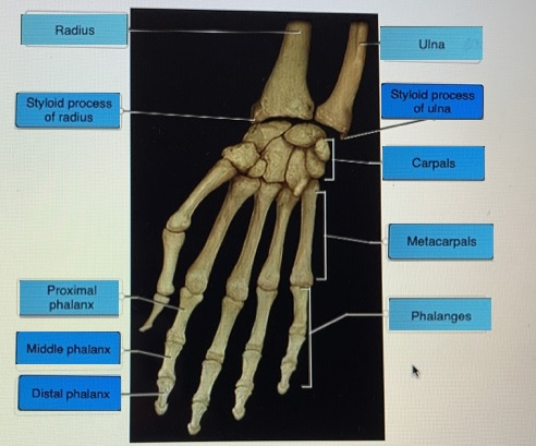
1. The wrist joint is made up of the radius, ulna, and carpal bones. This is composed of three main joints which include distal raioulnar joint, radiocarpal joint, and ulnocarpaljoints.
The hand is composed of multiple bones which include carpals, metacarpals, and phalanges. The phalanges are divided in to proximal, middle , and distal phalanges.
Add Answer to:
fill in the blank
Label the structures of the wrist and hand. Styloid process of ulna...
fill in the blank Label the structures of the bone. Proximal epiphysis Distal epiphysis Head Shaft...
 fill in the blank
Label the structures of the bone. Proximal epiphysis Distal epiphysis Head Shaft (diaphysis) Neck Femur Reset Zoom
fill in the blank
Label the structures of the bone. Proximal epiphysis Distal epiphysis Head Shaft (diaphysis) Neck Femur Reset Zoom
Label the structures of the ankle and foot. Tarsals Distal phalanx Middle phalanx Phalanges Proximal phalanx...
 Label the structures of the ankle and foot. Tarsals Distal phalanx Middle phalanx Phalanges Proximal phalanx Metatarsals
label the parts on the humerus: head,greater tubercle 4. Label the parts on the humerus: head,...
 label the parts on the humerus: head,greater tubercle
4. Label the parts on the humerus: head, greater tubercle, lesser tubercle, intertubercular sulcus, deltoid tuberosity, olecranon fossa, coronoid fossa, trochlea, capitulum, medial epicondyle, lateral epicondyle head ofd humerus head of humeus greater tubercle lesser tubercle -deltoid tuberosity deltold tuberosity coronoid fossa olecranon fossa lateral epicondyle medial epycondyle medial -lateral epvcondyle trochlea trochlea Anterior view Posterior view 5. Label the two bones located in the forearm and identify specific parts of their...
label the parts on the humerus: head,greater tubercle
4. Label the parts on the humerus: head, greater tubercle, lesser tubercle, intertubercular sulcus, deltoid tuberosity, olecranon fossa, coronoid fossa, trochlea, capitulum, medial epicondyle, lateral epicondyle head ofd humerus head of humeus greater tubercle lesser tubercle -deltoid tuberosity deltold tuberosity coronoid fossa olecranon fossa lateral epicondyle medial epycondyle medial -lateral epvcondyle trochlea trochlea Anterior view Posterior view 5. Label the two bones located in the forearm and identify specific parts of their...
fill in the blank Label the structures of the pelvis. Sacrum Ischium Intervertebral disc Pubis Coccyx...
 fill in the blank
Label the structures of the pelvis. Sacrum Ischium Intervertebral disc Pubis Coccyx llium Lumbar vertebra Reset Zoom
fill in the blank
Label the structures of the pelvis. Sacrum Ischium Intervertebral disc Pubis Coccyx llium Lumbar vertebra Reset Zoom
87- Chapter 5 The Skeletal System 22. Figure 5-10 is a diagram of the hand. Select...
 87- Chapter 5 The Skeletal System 22. Figure 5-10 is a diagram of the hand. Select different colors for the following structures, and use them to color the coding circles and the corresponding structures in the diagram Carpals O Metacarpals sive Phalanges 1000 Sphalanges - Metacarpas Carpals Radius Ulna Figure 5-10 23. Compare the pectoral and pelvic girdles by choosing descriptive terms from the key choices. Insert the appropriate key letters in the answer blanks. Key Choices A Flexibility D....
fill in the blank Label the structures of the bone using the hints provided. Sphenoid bone...
 fill in the blank
Label the structures of the bone using the hints provided. Sphenoid bone Foramen ovale Foramen rotundum Superior orbital fissure Superior orbital Posterior view fissure Foramen spinosum Foramen rotundum Foramen ovale Foramen spinosum Superior view Reset Zoom
fill in the blank
Label the structures of the bone using the hints provided. Sphenoid bone Foramen ovale Foramen rotundum Superior orbital fissure Superior orbital Posterior view fissure Foramen spinosum Foramen rotundum Foramen ovale Foramen spinosum Superior view Reset Zoom
label the structures of the right hand anterior view. Pectoral Girdle and Superior Appendages Label the...
 label the structures of the right hand anterior
view.
Pectoral Girdle and Superior Appendages Label the structures of the right hand anterior view Pisiform Lunate Triquetrum Trapezium Hamate Trapezoid Capitate Scaphoid Reset Zoom Ne 15 of 22 < Prev
Pectoral Girdle and Superior Appendages Label the structures of the right hand anterior view Pisiform Lunate Triquetrum Trapezium Hamate Trapezoid Capitate Scaphoid Reset Zoom Ne 15 of 22
label the structures of the right hand anterior
view.
Pectoral Girdle and Superior Appendages Label the structures of the right hand anterior view Pisiform Lunate Triquetrum Trapezium Hamate Trapezoid Capitate Scaphoid Reset Zoom Ne 15 of 22 < Prev
Pectoral Girdle and Superior Appendages Label the structures of the right hand anterior view Pisiform Lunate Triquetrum Trapezium Hamate Trapezoid Capitate Scaphoid Reset Zoom Ne 15 of 22
Pre-Lab #4 (This will help you prepare for the Skeletal Anatomy Test at end of Week...
Pre-Lab #4 (This will help you prepare for the Skeletal Anatomy Test at end of Week 5). Diagram (or obtain an unlabeled digital image of) and label the bones below. You can obtain unlabeled images online. For each bone complete the following tasks for Pre-Lab #4 Identify the bone and whether it is right or left (if that applies). Identify any markings or structures listed below on the bone (ex: fossa, cavity, process, etc). Identify any specific joints/articulations this bone...
fill in the blank Label the bones in the superior view of the cranial cavity. Frontal...
 fill in the blank
Label the bones in the superior view of the cranial cavity. Frontal bone Parietal bone Sphenoid bone Petrous portion Occipital bone Ethmoid bone Temporal bone Squamous portion Reset Zoom
fill in the blank
Label the bones in the superior view of the cranial cavity. Frontal bone Parietal bone Sphenoid bone Petrous portion Occipital bone Ethmoid bone Temporal bone Squamous portion Reset Zoom
fill in the blank Label the specific bony features of the superior skull. Skull Occipital bone...
 fill in the blank
Label the specific bony features of the superior skull. Skull Occipital bone Sutural bone DONE FEATURES Sagittal suture Parietal bone Frontal bone Coronal suture Lambdoid suture Superior view Reset Zoom
fill in the blank
Label the specific bony features of the superior skull. Skull Occipital bone Sutural bone DONE FEATURES Sagittal suture Parietal bone Frontal bone Coronal suture Lambdoid suture Superior view Reset Zoom
 fill in the blank
Label the structures of the bone. Proximal epiphysis Distal epiphysis Head Shaft (diaphysis) Neck Femur Reset Zoom
fill in the blank
Label the structures of the bone. Proximal epiphysis Distal epiphysis Head Shaft (diaphysis) Neck Femur Reset Zoom
 Label the structures of the ankle and foot. Tarsals Distal phalanx Middle phalanx Phalanges Proximal phalanx Metatarsals
 label the parts on the humerus: head,greater tubercle
4. Label the parts on the humerus: head, greater tubercle, lesser tubercle, intertubercular sulcus, deltoid tuberosity, olecranon fossa, coronoid fossa, trochlea, capitulum, medial epicondyle, lateral epicondyle head ofd humerus head of humeus greater tubercle lesser tubercle -deltoid tuberosity deltold tuberosity coronoid fossa olecranon fossa lateral epicondyle medial epycondyle medial -lateral epvcondyle trochlea trochlea Anterior view Posterior view 5. Label the two bones located in the forearm and identify specific parts of their...
label the parts on the humerus: head,greater tubercle
4. Label the parts on the humerus: head, greater tubercle, lesser tubercle, intertubercular sulcus, deltoid tuberosity, olecranon fossa, coronoid fossa, trochlea, capitulum, medial epicondyle, lateral epicondyle head ofd humerus head of humeus greater tubercle lesser tubercle -deltoid tuberosity deltold tuberosity coronoid fossa olecranon fossa lateral epicondyle medial epycondyle medial -lateral epvcondyle trochlea trochlea Anterior view Posterior view 5. Label the two bones located in the forearm and identify specific parts of their...
 fill in the blank
Label the structures of the pelvis. Sacrum Ischium Intervertebral disc Pubis Coccyx llium Lumbar vertebra Reset Zoom
fill in the blank
Label the structures of the pelvis. Sacrum Ischium Intervertebral disc Pubis Coccyx llium Lumbar vertebra Reset Zoom
 87- Chapter 5 The Skeletal System 22. Figure 5-10 is a diagram of the hand. Select different colors for the following structures, and use them to color the coding circles and the corresponding structures in the diagram Carpals O Metacarpals sive Phalanges 1000 Sphalanges - Metacarpas Carpals Radius Ulna Figure 5-10 23. Compare the pectoral and pelvic girdles by choosing descriptive terms from the key choices. Insert the appropriate key letters in the answer blanks. Key Choices A Flexibility D....
 fill in the blank
Label the structures of the bone using the hints provided. Sphenoid bone Foramen ovale Foramen rotundum Superior orbital fissure Superior orbital Posterior view fissure Foramen spinosum Foramen rotundum Foramen ovale Foramen spinosum Superior view Reset Zoom
fill in the blank
Label the structures of the bone using the hints provided. Sphenoid bone Foramen ovale Foramen rotundum Superior orbital fissure Superior orbital Posterior view fissure Foramen spinosum Foramen rotundum Foramen ovale Foramen spinosum Superior view Reset Zoom
 label the structures of the right hand anterior
view.
Pectoral Girdle and Superior Appendages Label the structures of the right hand anterior view Pisiform Lunate Triquetrum Trapezium Hamate Trapezoid Capitate Scaphoid Reset Zoom Ne 15 of 22 < Prev
Pectoral Girdle and Superior Appendages Label the structures of the right hand anterior view Pisiform Lunate Triquetrum Trapezium Hamate Trapezoid Capitate Scaphoid Reset Zoom Ne 15 of 22
label the structures of the right hand anterior
view.
Pectoral Girdle and Superior Appendages Label the structures of the right hand anterior view Pisiform Lunate Triquetrum Trapezium Hamate Trapezoid Capitate Scaphoid Reset Zoom Ne 15 of 22 < Prev
Pectoral Girdle and Superior Appendages Label the structures of the right hand anterior view Pisiform Lunate Triquetrum Trapezium Hamate Trapezoid Capitate Scaphoid Reset Zoom Ne 15 of 22
 fill in the blank
Label the bones in the superior view of the cranial cavity. Frontal bone Parietal bone Sphenoid bone Petrous portion Occipital bone Ethmoid bone Temporal bone Squamous portion Reset Zoom
fill in the blank
Label the bones in the superior view of the cranial cavity. Frontal bone Parietal bone Sphenoid bone Petrous portion Occipital bone Ethmoid bone Temporal bone Squamous portion Reset Zoom
 fill in the blank
Label the specific bony features of the superior skull. Skull Occipital bone Sutural bone DONE FEATURES Sagittal suture Parietal bone Frontal bone Coronal suture Lambdoid suture Superior view Reset Zoom
fill in the blank
Label the specific bony features of the superior skull. Skull Occipital bone Sutural bone DONE FEATURES Sagittal suture Parietal bone Frontal bone Coronal suture Lambdoid suture Superior view Reset Zoom
Most questions answered within 3 hours.
-
Which food law was passed in 1996 and changed how pesticide
residues on food were regulated...
asked 8 minutes ago -
companies either hire outside programmers to
write_____ software or use their own internal developers.
asked 7 minutes ago -
A magnetic dipole m(t) = m_0*cos(ωt) can be
described as current density j(r,t) = −cm(t) ×...
asked 7 minutes ago -
Probabilities and Counting. Yahtzee is a game that involves six
fair dice. When rolling all six...
asked 9 minutes ago -
What percent of revenue does net income represent for each
year?
Total Revenue
2017 = 60,319,000...
asked 31 minutes ago -
For Ti+2 (Z=22). Determine the correct ground state
& # of microstates. Use the correct tanabe...
asked 34 minutes ago -
Why did so many investment banks have to start buying CDO’s and
other mortgaged backed securities...
asked 49 minutes ago -
The mean cost of domestic airfares in the United States rose to
an all-time high of...
asked 1 hour ago -
1.Magazine Luiza is a Brazilian retail chain for consumer
electronics. The company currently has 100 stores...
asked 59 minutes ago -
What is the molarity of ZnCl2 that forms when 25.0 g of zinc
completely reacts with...
asked 1 hour ago -
For independent X and Y, we have probability density function
for them where pdf of X...
asked 1 hour ago -
The decomposition of SO2Cl2 is first order in SO2Cl2 and has a
rate constant of 1.42...
asked 1 hour ago