
Homework Answers


 The
pleura consists of:
The
pleura consists of:
1.Outer parietal peritoneum
2. Inner visceral peritoneum
3. Middle pleural cavity
So, Mark accordingly as picture is blurred at zoom. All these 3 makes pleura.
Add Answer to:
cartilage is the tiny corniculate cartilage. The artenoid and corniculate carties are paired with the are...
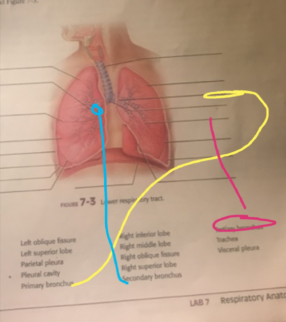
trachea primary bronchi plurae FIGURE 7-3 Lower respiratory tract. TERMS FOR FIGURE 7-3 Cardiac notch Carina...
 trachea primary bronchi plurae FIGURE 7-3 Lower respiratory tract. TERMS FOR FIGURE 7-3 Cardiac notch Carina Diaphragm Horizontal fissure Left inferior lobe Left oblique fissure Left superior lobe Parietal pleura Pleural cavity Primary bronchus Right inferior lobe Right middle lobe Right oblique fissure Right superior lobe Secondary bronchus Tertiary bronchus Trachea Visceral pleura
(2.5 pts 2. Within the nasal cavity, posterior to the vestibule are three bony folds that...
 (2.5 pts 2. Within the nasal cavity, posterior to the vestibule are three bony folds that project from the lateral walls toward the septum. O The vibrissae The perpendicular plate of the ethmoid The nasal conchae The choanae O 3. Which region of the pharynx is found between the soft palate and the top of the epiglottis? 2.5 pts O Nasopharynx Oropharynx Laryngopharynx O Suprapharynx 4. Which cartilage has an anterior peak the laryngeal prominence also known as the "Adam's...
Art-Labeling Activity: The Pleurae and Pleural Cavities Part A Drag the appropriate labels to their respective...
 Art-Labeling Activity: The Pleurae and Pleural Cavities Part A Drag the appropriate labels to their respective targets. Reset Help Pleural cavity with serous fluid Vertebra Pericardial cavity Left lung Heart Left oblique fissure Horizontal fissure Parietal pleura Visceral pleura Right lung Anterior Inferior view Posterior -Thoracic wall
D 1. These are peripheral chemoreceptors that monitor changes in respiratory gasses and blood acidity levels...
 D 1. These are peripheral chemoreceptors that monitor changes in respiratory gasses and blood acidity levels carotid bodies medulla oblongata vagus nerve D 2. The region of the lung served by secondary, or lobar, bronchi. lobule bronchopulmonary segment lobe D 3. The respiratory zone begins at the large bronchioles. False True D 4. The serous membrane lining of the wall of the thoracic cavity. parietal pericardium visceral pleura parietal pleura visceral pericardium D 5. The smallest airway of the bronchial...
10. The Respiratory System A. Anatomy of the respiratory system 1. Label the parts of the...
 10. The Respiratory System A. Anatomy of the respiratory system 1. Label the parts of the upper respiratory system: cente, epiglottis, external naris, laryngopharynx, nasal cavity, nasopharynx, lingual tonsil, opening of eustachian tube, oropharynx, palatine tonsil, thyroid cartilage, trachea, vocal folds, pharyngeal tonsil, nasal vestibule Opening of eustechian tube Oral cavity- Esophagus 113 2. Label the parts of the lower respiratory system: epiglottis, inferior lobe of left lung, inferior lobe of right lung, larynx, middle lobe of right lung, primary...
pre-lab exercise 21-1 Name Section Date PRE-LAB EXERCISES Complete the following esxercises prior to coming to...
 pre-lab exercise 21-1
Name Section Date PRE-LAB EXERCISES Complete the following esxercises prior to coming to lab, using your textbook and lab manvual for seference Pre-Lab Exercise 21-1 Key Terms You should be familiar with the following terms before coming to lab. Term General Structures of the Respiratory System Respiratory tract finition Parietal pleura Visceral pleura Pleural cavity Lungs and lobes Structures of the Respiratory Tract Nasal cavity Pharynx 21 Larynx Trachea Primary bronchi Secondary bronchi 510 I Exploring Anatomy...
pre-lab exercise 21-1
Name Section Date PRE-LAB EXERCISES Complete the following esxercises prior to coming to lab, using your textbook and lab manvual for seference Pre-Lab Exercise 21-1 Key Terms You should be familiar with the following terms before coming to lab. Term General Structures of the Respiratory System Respiratory tract finition Parietal pleura Visceral pleura Pleural cavity Lungs and lobes Structures of the Respiratory Tract Nasal cavity Pharynx 21 Larynx Trachea Primary bronchi Secondary bronchi 510 I Exploring Anatomy...
I just need help finishing up this diagram question. WE 3. Sketch the respiratory system including...
 I just need help finishing up this diagram question.
WE 3. Sketch the respiratory system including the following elements: left lung, right lung, trachea, right main bronchus, left main bronchus, bronchioles, alveoli, diaphragm, pleural sac, pleura, bronchi, pulmonary capillaries, pulmonary artery, arteriole, pulmonary vein, and veinule. Left Lung Right Main aconchus Trachea Right Lung Bronchioles Left Main Siem Bronchus -Bronchi & Pleura & Diaphragm
I just need help finishing up this diagram question.
WE 3. Sketch the respiratory system including the following elements: left lung, right lung, trachea, right main bronchus, left main bronchus, bronchioles, alveoli, diaphragm, pleural sac, pleura, bronchi, pulmonary capillaries, pulmonary artery, arteriole, pulmonary vein, and veinule. Left Lung Right Main aconchus Trachea Right Lung Bronchioles Left Main Siem Bronchus -Bronchi & Pleura & Diaphragm
Answer these correctly please, thanks! LAB REPORT 31 Nam Respiratory Structures Section checklist for your study...
 Answer these correctly please, thanks!
LAB REPORT 31 Nam Respiratory Structures Section checklist for your study of the human model and the shore luck. Use your textbook for help with the this tableasach that asks for functions. Pluck Structure Functions) Naual cavity Nasal septum Nes internal, external Hard palate Sart palate Cache: superior, middle inferior Purnal sinuses: maxillary. frontal, ethmoid, sphenoid Nesopharynx Oropharynx Laryngopharynx Laryngeal cartilages Vestibular folds TOODOO O OOOOOOOOOOOOOOOOO OOOOOOOOOOOOOOOOOOOOOOOO Vocal folds Trachea Primary bronchi Secondary bronchi Tertiary...
Answer these correctly please, thanks!
LAB REPORT 31 Nam Respiratory Structures Section checklist for your study of the human model and the shore luck. Use your textbook for help with the this tableasach that asks for functions. Pluck Structure Functions) Naual cavity Nasal septum Nes internal, external Hard palate Sart palate Cache: superior, middle inferior Purnal sinuses: maxillary. frontal, ethmoid, sphenoid Nesopharynx Oropharynx Laryngopharynx Laryngeal cartilages Vestibular folds TOODOO O OOOOOOOOOOOOOOOOO OOOOOOOOOOOOOOOOOOOOOOOO Vocal folds Trachea Primary bronchi Secondary bronchi Tertiary...
Drag the labels onto the diagram to identify the anatomical features of the right lung (lateral surface). Reset Ape...
 Drag the labels onto the diagram to identify the anatomical features of the right lung (lateral surface). Reset Apex Base Horizontal fissure Oblique fissure Middle lobe Superior lobe Inferior lobe
Drag the labels onto the diagram to identify the anatomical features of the right lung (lateral surface). Reset Apex Base Horizontal fissure Oblique fissure Middle lobe Superior lobe Inferior lobe
Drag the labels onto the diagram to identify the anatomical features of the right lung (lateral surface). Reset Apex Base Horizontal fissure Oblique fissure Middle lobe Superior lobe Inferior lobe
Instructors may of the Review SH using Masterin REVIEW SHEET Anatomy of the Respiratory System EXERCISE...
 Instructors may of the Review SH using Masterin REVIEW SHEET Anatomy of the Respiratory System EXERCISE Name Lab Time/Date Upper and lower Respiratory System Structures 1. Complete the labeling of the model of the respiratory structures (sagittal section) shown below. 2. Two pairs of mucosal folds are found in the larynx. Which pair are the true vocal cords (superior or infer 3. Name the specific cartilages in the larynx that correspond to the following descriptions. shaped like a ring forms...
 trachea primary bronchi plurae FIGURE 7-3 Lower respiratory tract. TERMS FOR FIGURE 7-3 Cardiac notch Carina Diaphragm Horizontal fissure Left inferior lobe Left oblique fissure Left superior lobe Parietal pleura Pleural cavity Primary bronchus Right inferior lobe Right middle lobe Right oblique fissure Right superior lobe Secondary bronchus Tertiary bronchus Trachea Visceral pleura
 (2.5 pts 2. Within the nasal cavity, posterior to the vestibule are three bony folds that project from the lateral walls toward the septum. O The vibrissae The perpendicular plate of the ethmoid The nasal conchae The choanae O 3. Which region of the pharynx is found between the soft palate and the top of the epiglottis? 2.5 pts O Nasopharynx Oropharynx Laryngopharynx O Suprapharynx 4. Which cartilage has an anterior peak the laryngeal prominence also known as the "Adam's...
 Art-Labeling Activity: The Pleurae and Pleural Cavities Part A Drag the appropriate labels to their respective targets. Reset Help Pleural cavity with serous fluid Vertebra Pericardial cavity Left lung Heart Left oblique fissure Horizontal fissure Parietal pleura Visceral pleura Right lung Anterior Inferior view Posterior -Thoracic wall
 D 1. These are peripheral chemoreceptors that monitor changes in respiratory gasses and blood acidity levels carotid bodies medulla oblongata vagus nerve D 2. The region of the lung served by secondary, or lobar, bronchi. lobule bronchopulmonary segment lobe D 3. The respiratory zone begins at the large bronchioles. False True D 4. The serous membrane lining of the wall of the thoracic cavity. parietal pericardium visceral pleura parietal pleura visceral pericardium D 5. The smallest airway of the bronchial...
 10. The Respiratory System A. Anatomy of the respiratory system 1. Label the parts of the upper respiratory system: cente, epiglottis, external naris, laryngopharynx, nasal cavity, nasopharynx, lingual tonsil, opening of eustachian tube, oropharynx, palatine tonsil, thyroid cartilage, trachea, vocal folds, pharyngeal tonsil, nasal vestibule Opening of eustechian tube Oral cavity- Esophagus 113 2. Label the parts of the lower respiratory system: epiglottis, inferior lobe of left lung, inferior lobe of right lung, larynx, middle lobe of right lung, primary...
 pre-lab exercise 21-1
Name Section Date PRE-LAB EXERCISES Complete the following esxercises prior to coming to lab, using your textbook and lab manvual for seference Pre-Lab Exercise 21-1 Key Terms You should be familiar with the following terms before coming to lab. Term General Structures of the Respiratory System Respiratory tract finition Parietal pleura Visceral pleura Pleural cavity Lungs and lobes Structures of the Respiratory Tract Nasal cavity Pharynx 21 Larynx Trachea Primary bronchi Secondary bronchi 510 I Exploring Anatomy...
pre-lab exercise 21-1
Name Section Date PRE-LAB EXERCISES Complete the following esxercises prior to coming to lab, using your textbook and lab manvual for seference Pre-Lab Exercise 21-1 Key Terms You should be familiar with the following terms before coming to lab. Term General Structures of the Respiratory System Respiratory tract finition Parietal pleura Visceral pleura Pleural cavity Lungs and lobes Structures of the Respiratory Tract Nasal cavity Pharynx 21 Larynx Trachea Primary bronchi Secondary bronchi 510 I Exploring Anatomy...
 I just need help finishing up this diagram question.
WE 3. Sketch the respiratory system including the following elements: left lung, right lung, trachea, right main bronchus, left main bronchus, bronchioles, alveoli, diaphragm, pleural sac, pleura, bronchi, pulmonary capillaries, pulmonary artery, arteriole, pulmonary vein, and veinule. Left Lung Right Main aconchus Trachea Right Lung Bronchioles Left Main Siem Bronchus -Bronchi & Pleura & Diaphragm
I just need help finishing up this diagram question.
WE 3. Sketch the respiratory system including the following elements: left lung, right lung, trachea, right main bronchus, left main bronchus, bronchioles, alveoli, diaphragm, pleural sac, pleura, bronchi, pulmonary capillaries, pulmonary artery, arteriole, pulmonary vein, and veinule. Left Lung Right Main aconchus Trachea Right Lung Bronchioles Left Main Siem Bronchus -Bronchi & Pleura & Diaphragm
 Answer these correctly please, thanks!
LAB REPORT 31 Nam Respiratory Structures Section checklist for your study of the human model and the shore luck. Use your textbook for help with the this tableasach that asks for functions. Pluck Structure Functions) Naual cavity Nasal septum Nes internal, external Hard palate Sart palate Cache: superior, middle inferior Purnal sinuses: maxillary. frontal, ethmoid, sphenoid Nesopharynx Oropharynx Laryngopharynx Laryngeal cartilages Vestibular folds TOODOO O OOOOOOOOOOOOOOOOO OOOOOOOOOOOOOOOOOOOOOOOO Vocal folds Trachea Primary bronchi Secondary bronchi Tertiary...
Answer these correctly please, thanks!
LAB REPORT 31 Nam Respiratory Structures Section checklist for your study of the human model and the shore luck. Use your textbook for help with the this tableasach that asks for functions. Pluck Structure Functions) Naual cavity Nasal septum Nes internal, external Hard palate Sart palate Cache: superior, middle inferior Purnal sinuses: maxillary. frontal, ethmoid, sphenoid Nesopharynx Oropharynx Laryngopharynx Laryngeal cartilages Vestibular folds TOODOO O OOOOOOOOOOOOOOOOO OOOOOOOOOOOOOOOOOOOOOOOO Vocal folds Trachea Primary bronchi Secondary bronchi Tertiary...
 Drag the labels onto the diagram to identify the anatomical features of the right lung (lateral surface). Reset Apex Base Horizontal fissure Oblique fissure Middle lobe Superior lobe Inferior lobe
Drag the labels onto the diagram to identify the anatomical features of the right lung (lateral surface). Reset Apex Base Horizontal fissure Oblique fissure Middle lobe Superior lobe Inferior lobe
Drag the labels onto the diagram to identify the anatomical features of the right lung (lateral surface). Reset Apex Base Horizontal fissure Oblique fissure Middle lobe Superior lobe Inferior lobe
 Instructors may of the Review SH using Masterin REVIEW SHEET Anatomy of the Respiratory System EXERCISE Name Lab Time/Date Upper and lower Respiratory System Structures 1. Complete the labeling of the model of the respiratory structures (sagittal section) shown below. 2. Two pairs of mucosal folds are found in the larynx. Which pair are the true vocal cords (superior or infer 3. Name the specific cartilages in the larynx that correspond to the following descriptions. shaped like a ring forms...
Most questions answered within 3 hours.
-
if the plasmid concentration is 100 ng/ul how much ul need to be
added to the...
asked 6 minutes ago -
I need this code in java.
Loops do just what they sound like they should -...
asked 3 minutes ago -
Your company wants to raise $11.0 million by issuing 30-year
zero-coupon bonds. If the yield to...
asked 27 minutes ago -
Determine the pressure exerted by 3.99 mol of gas in a 2.92-L
container at 32oC
asked 6 minutes ago -
Riverbed Corp purchased a new blending machine for $3,050.01. It
paid $520.22 down and financed the...
asked 16 minutes ago -
Explain what is involved in pursuing a policy of
nondiscrimination.
asked 9 minutes ago -
A company faces an inverse demand curve of p = 17 − 2Q and its
cost...
asked 23 minutes ago -
The number of claims for lost luggage in a small city airport
averages 8 per day....
asked 24 minutes ago -
Radio waves, from your favorite radio station has a frequency of
( 89.3) MHz (megahertz). What...
asked 28 minutes ago -
In how many ways can one obtain at least three jacks in a six
card hand?...
asked 41 minutes ago -
Neal Nicely was driving on the parkway when he noticed two little
2. old ladies on...
asked 47 minutes ago -
The size of a movement ALONG a demand curve caused by a shift in
supply would...
asked 43 minutes ago