
Homework Answers
Ans 1-: Construction of mRNA from DNA is called transcription and synthesis of amino acid sequence from mRNA is called translation.
TRANSCRIPTION
Transcription is the process by which DNA is copied (transcribed) to mRNA, which carries the information needed for protein synthesis. Transcription takes place in two broad steps. First, pre-messenger RNA is formed, with the involvement of RNA polymerase enzymes. The process relies on Watson-Crick base pairing, and the resultant single strand of RNA is the reverse-complement of the original DNA sequence. The pre-messenger RNA is then "edited" to produce the desired mRNA molecule in a process called RNA splicing.
Formation of pre-messenger RNA
The mechanism of transcription has parallels in that of DNA replication. As with DNA replication, partial unwinding of the double helix must occur before transcription can take place, and it is the RNA polymerase enzymes that catalyze this process.
Unlike DNA replication, in which both strands are copied, only one strand is transcribed. The strand that contains the gene is called the sense strand, while the complementary strand is the antisensestrand. The mRNA produced in transcription is a copy of the sense strand, but it is the antisense strand that is transcribed.
Ribonucleoside triphosphates (NTPs) align along the antisense DNA strand, with Watson-Crick base pairing (A pairs with U). RNA polymerase joins the ribonucleotides together to form a pre-messenger RNA molecule that is complementary to a region of the antisense DNA strand. Transcription ends when the RNA polymerase enzyme reaches a triplet of bases that is read as a "stop" signal. The DNA molecule re-winds to re-form the double helix.

Figure-:Transcription representation of the formation of pre-messenger RNA (orange) from double-stranded DNA (blue) in transcription.
RNA splicing
The pre-messenger RNA thus formed contains introns which are not required for protein synthesis. The pre-messenger RNA is chopped up to remove the introns and create messenger RNA (mRNA) in a process called RNA splicing

Figure-:RNA splicing Introns are spliced from the pre-messenger RNA to give messenger RNA (mRNA).
Alternative splicing
In alternative splicing, individual exons are either spliced or included, giving rise to several different possible mRNA products. Each mRNA product codes for a different protein isoform; these protein isoforms differ in their peptide sequence and therefore their biological activity. It is estimated that up to 60% of human gene products undergo alternative splicing.

Figure-:Alternative splicing- different mechanisms of alternative splicing exist − a cassette exon can be either included in or excluded from the final RNA (top), or two cassette exons may be mutually exclusive (bottom).
Alternative splicing contributes to protein diversity − a single gene transcript (RNA) can have thousands of different splicing patterns, and will therefore code for thousands of different proteins: a diverse proteome is generated from a relatively limited genome. Splicing is important in genetic regulation (alteration of the splicing pattern in response to cellular conditions changes protein expression). Perhaps not surprisingly, abnormal splicing patterns can lead to disease states including cancer.
Reverse transcription
In reverse transcription, RNA is "reverse transcribed" into DNA. This process, catalyzed by reverse transcriptase enzymes, allows retroviruses, including the human immunodeficiency virus (HIV), to use RNA as their genetic material. Reverse transcriptase enzymes have also found applications in biotechnology, allowing scientists to convert RNA to DNA for techniques such as PCR.
TRANSLATION
The mRNA formed in transcription is transported out of the nucleus, into the cytoplasm, to the ribosome (the cell's protein synthesis factory). Here, it directs protein synthesis. Messenger RNA is not directly involved in protein synthesis − transfer RNA (tRNA) is required for this. The process by which mRNA directs protein synthesis with the assistance of tRNA is called translation.
The ribosome is a very large complex of RNA and protein molecules. Each three-base stretch of mRNA (triplet) is known as a codon, and one codon contains the information for a specific amino acid. As the mRNA passes through the ribosome, each codon interacts with the anticodon of a specific transfer RNA (tRNA) molecule by Watson-Crick base pairing. This tRNA molecule carries an amino acid at its 3′-terminus, which is incorporated into the growing protein chain. The tRNA is then expelled from the ribosome. Figure shows the steps involved in protein synthesis.

Figure -:Translation(a) and (b) tRNA molecules bind to the two binding sites of the ribosome, and by hydrogen bonding to the mRNA; (c) a peptide bond forms between the two amino acids to make a dipeptide, while the tRNA molecule is left uncharged; (d) the uncharged tRNA molecule leaves the ribosome, while the ribosome moves one codon to the right (the dipeptide is translocated from one binding site to the other); (e) another tRNA molecule binds; (f) a peptide bond forms between the two amino acids to make a tripeptide; (g) the uncharged tRNA molecule leaves the ribosome.
TRANSFER RNA
Transfer RNA adopts a well defined tertiary structure which is normally represented in two dimensions as a cloverleaf shape, as in Figure 7. The structure of tRNA is shown in more detail in Figure 8.

Figure -: Two-dimensional structures of tRNA (transfer RNA)In some tRNAs the DHU arm has only three base pairs.
Each amino acid has its own special tRNA (or set of tRNAs). For example, the tRNA for phenylalanine (tRNAPhe) is different from that for histidine (tRNAHis). Each amino acid is attached to its tRNA through the 3′-OH group to form an ester which reacts with the α-amino group of the terminal amino-acid of the growing protein chain to form a new amide bond (peptide bond) during protein synthesis (Figure 9). The reaction of esters with amines is generally favourable but the rate of reaction is increased greatly in the ribosome.
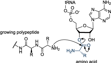
Figure - :Proteins of the growing polypeptide chain with the 3′-end of the charged tRNA. The amino acid is transferred from the tRNA molecule to the protein.
Each transfer RNA molecule has a well defined tertiary structure that is recognized by the enzyme aminoacyl tRNA synthetase, which adds the correct amino acid to the 3′-end of the uncharged tRNA. The presence of modified nucleosides is important in stabilizing the tRNA structure. Some of these modifications are shown in Figure
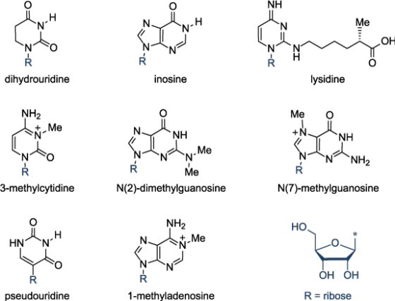
Figure | Modified bases in tRNAStructures of some of the modified bases found in tRNA.
THE GENETIC CODE
The genetic code is almost universal. It is the basis of the transmission of hereditary information by nucleic acids in all organisms. There are four bases in RNA (A,G,C and U), so there are 64 possible triplet codes (43 = 64). In theory only 22 codes are required: one for each of the 20 naturally occurring amino acids, with the addition of a start codon and a stop codon (to indicate the beginning and end of a protein sequence). Many amino acids have several codes (degeneracy), so that all 64 possible triplet codes are used. For example Arg and Ser each have 6 codons whereas Trp and Met have only one. No two amino acids have the same code but amino acids whose side-chains have similar physical or chemical properties tend to have similar codon sequences, e.g. the side-chains of Phe, Leu, Ile, Val are all hydrophobic, and Asp and Glu are both carboxylic acids (see Figure 11). This means that if the incorrect tRNA is selected during translation (owing to mispairing of a single base at the codon-anticodon interface) the misincorporated amino acid will probably have similar properties to the intended tRNA molecule. Although the resultant protein will have one incorrect amino acid it stands a high probability of being functional. Organisms show "codon bias" and use certain codons for a particular amino acid more than others. For example, the codon usage in humans is different from that in bacteria; it can sometimes be difficult to express a human protein in bacteria because the relevant tRNA might be present at too low a concentration.

Figure -: The Genetic code − triplet codon assignments for the 20 amino acids. As well as coding for methionine, AUG is used as a start codon, initiating protein biosynthesis.
Ans 2-:
DNA REPLICATION
Each time a cell divides, each of its double strands of DNA splits into two single strands. Each of these single strands acts as a template for a new strand of complementary DNA. As a result, each new cell has its own complete genome. This process is known as DNA replication. Replication is controlled by the Watson-Crick pairing of the bases in the template strand with incoming deoxynucleoside triphosphates, and is directed by DNA polymerase enzymes. It is a complex process, particularly in eukaryotes, involving an array of enzymes. A simplified version of bacterial DNA replication is described in Figure

Figure | DNA replication in bacteriaSimplified representation of DNA replication in bacteria.
DNA biosynthesis proceeds in the 5′- to 3′-direction. This makes it impossible for DNA polymerases to synthesize both strands simultaneously. A portion of the double helix must first unwind, and this is mediated by helicase enzymes.
The leading strand is synthesized continuously but the opposite strand is copied in short bursts of about 1000 bases, as the lagging strand template becomes available. The resulting short strands are called Okazaki fragments (after their discoverers, Reiji and Tsuneko Okazaki). Bacteria have at least three distinct DNA polymerases: Pol I, Pol II and Pol III; it is Pol III that is largely involved in chain elongation. Strangely, DNA polymerases cannot initiate DNA synthesis de novo, but require a short primer with a free 3′-hydroxyl group. This is produced in the lagging strand by an RNA polymerase (called DNA primase) that is able to use the DNA template and synthesize a short piece of RNA around 20 bases in length. Pol III can then take over, but it eventually encounters one of the previously synthesized short RNA fragments in its path. At this point Pol I takes over, using its 5′- to 3′-exonuclease activity to digest the RNA and fill the gap with DNA until it reaches a continuous stretch of DNA. This leaves a gap between the 3′-end of the newly synthesized DNA and the 5′-end of the DNA previously synthesized by Pol III. The gap is filled by DNA ligase, an enzyme that makes a covalent bond between a 5′-phosphate and a 3′-hydroxyl group (Figure 3). The initiation of DNA replication at the leading strand is more complex and is discussed in detail in more specialized texts.
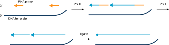
Figure | DNA polymerases in DNA replicationSimplified representation of the action of DNA polymerases in DNA replication in bacteria.
Mistakes in DNA replication
DNA replication is not perfect. Errors occur in DNA replication, when the incorrect base is incorporated into the growing DNA strand. This leads to mismatched base pairs, or mispairs. DNA polymerases have proofreading activity, and a DNA repair enzymes have evolved to correct these mistakes. Occasionally, mispairs survive and are incorporated into the genome in the next round of replication. These mutations may have no consequence, they may result in the death of the organism, they may result in a genetic disease or cancer; or they may give the organism a competitive advantage over its neighbours, which leads to evolution by natural selection.
Ans 3-: Packaging of DNA-:
The double helix of DNA is highly negatively charged due to all
the negatively charged phosphates in the backbone. All that
negative charge must be counterbalanced by a positive charge, and
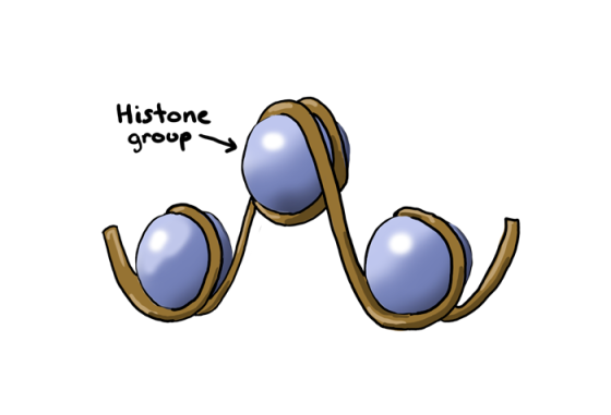
the cell makes proteins called histones that bind DNA and aid in
DNA's packaging. Histones are positively charged proteins that wrap
up DNA through interactions between their positive charges and the
negative charges of DNA. Double-stranded DNA loops around 8
histones twice, forming the nucleosome, which is the building block
of chromatin packaging.

DNA can be further packaged by forming coils of nucleosomes, called
chromatin fibers. These fibers are condensed into chromosomes
during mitosis, or the process of cell division. However, packaging
of chromatin into chromosomes that we are most familiar with occurs
only during a few stages of mitosis. Most of the time, DNA is
loosely packaged.
Histones are positively charged proteins that facilitate the
packing of DNA into condensed chromatin fibers. They are basically
the TupperwareTM of DNA packaging, and they come in many
kitchen-friendly colors. Histones have many arginine and lysine
amino acids that easily bind to the negatively charged DNA, based
on Paula Abdul's principle that opposites attract. Just kidding on
that last part. DNA is highly negatively charged because of the
phosphate group of each nucleotide is negatively charged.
Histones are divided into two groups:
- Core histones
- Linker histones
Core histones are H2A, H2B, H3, and H4, where two H3/H4 dimers
(H3 and H4 hooked together) and two H2A/H2B dimers (these two
hooked together) form the octamer (all eight of these guys
together). Linker histone H1 basically locks the DNA in place onto
the nucleosome and can be removed for transcriptionwhile linker
histone H5 is a variant of H1 predominantly used in birds.

H1, H2A, H2B, H3, H4, and H5 are all names that define families of
proteins. Individual histone proteins are specific for certain
types of DNA or certain cell types. Just as H5 is the avian version
of H1, there are individual histone proteins that package certain
regions of DNA, or package DNA in specific tissue types. Just like
you would not put a giant pot of chili in small Tupperware
containers (or maybe you would…we try not to judge, but
seriously?), specific histones are important for specific parts of
DNA.
One important aspect of histones is that they can be changed to
alter how much packing the DNA is capable of. There are several
modifications that affect how well DNA is packaged. The three major
types of modifications can be seen in the following table.
| Modification | Modification Structure (R = chemical functional group) | Charge | Effect |
| Methylation | R-CH3 | Neutral | Increases packing |
| Acetylation | R-COCH3 | Negative | Decreases packing |
| Phosphorylation | R-PO4 | Negative | Decreases packing |
Normally, histones are positively charged molecules, and the
addition of methyl groups (methylation) makes them more hydrophobic
(water-hating). Hydrophobic molecules tend to stick together, and
increasing histone methylation will cause the histones to pack even
more tightly than usual.
Acetylation (adding an acetyl group) and phosphorylation (adding a
phosphate group) make the histones more negatively charged because
acetyl and phosphoryl groups are negative. They are "glass is half
empty" molecules. By making histones more negatively charged, their
grip on DNA will be much looser because DNA is also negatively
charged. Similar charges (negative and negative) repel one
another.
One of the perks of packaging DNA is that you can separate it
into things you use a lot and things you do not. Unless you are a
maniacal hoarder, every fall, you put away your summer clothes for
things more winter-appropriate. In the same way, certain parts of
DNA are only important for certain times. However, some things you
need year-round, like shoes, so there is no point in putting those
things away. The cell does the same thing with DNA.
Regions that are necessary for making proteins and are important
for the cell are loosely packed and called euchromatin. By having a
loose packing of DNA in euchromatin, proteins involved in
transcription can easily get in and make RNA (see Genes to Proteins
section for more detail). On the other hand, some regions of DNA
you do not need except for special occasions, like that velvet suit
you have that you never wear. These regions are called
heterochromatin and are tightly packed through DNA as well as
through good ol' histone methylation.

Enzymes that add acetyl groups to histones are called histone
acetyltransferases (HATs) while those that remove acetyl groups are
called histone deacetylases (HDACs). Enzymes that add methyl groups
are called histone methyltransferases (HMTs). Activity of these
enzymes affects whether or not regions of DNA are tightly packed,
and unable to transcribe, or are loosely packed and therefore,
highly transcribed.
Histone methylation is a tricky concept, though, because usually,
histone methylation goes along with methylation of cytosines in
DNA, called DNA methylation. Together, these processes create
regions of DNA that cannot be transcribed. However, sometimes,
methylation of positively charged amino acids in histones promotes
transcriptional activation, but only when DNA is not methylated.
The methylation of DNA and modifications of histones that affect
transcription are the focus of study called epigenetics.
When the cell is undergoing the process of mitosis, chromatin
packing is important, and this packing is done by packing DNA into
condensed chromatin fibers to the point where they are the
recognized chromosomes that we know and love…or "really like," if
you are unprepared to make that kind of commitment. These
chromosomes divide into daughter cells, and after mitosis is
complete, the DNA is unpacked so transcription can occur again.
Therefore, we can think of mitosis like a big DNA moving day. The
packing starts with HDACs and HMTs tightening the packaging, and
once mitosis is completed, HATs and phosphoryltransferases (HPTs)
reduce the packaging.
Add Answer to:
and mRNA uence (yae Car ue a chart o illstra te escripthon le yding strand and steps is her DVA he puctaqit -nin and mRNA uence (yae Car ue a chart o illstra te escripthon le yding strand and...
2. Car Class ak tor the imake of theca speed o he a rent sqeedl The Car í should h e an intai it ...
 2. Car Class ak tor the imake of theca speed o he a rent sqeedl The Car í should h e an intai it tha that aitt ti ii inn make as argus, Thes va oald te ta - 瞬 The dlas shond e hawe the tloi tho! acce:erste brak The brake method beeld subtra s trom de paed dtdt get eed The getuspend metho eild ne teuts Next, deign a program that carm r b trt times. After cach...
ice cream. She asks 100 of her regular customers te he best. The results are shown...
 ice cream. She asks 100 of her regular customers te he best. The results are shown in the follo taie test and pick the navour they like tahle 7(1 mi) Quinn's Cafe serves i Green tea 2 Lemon Caffee Total 100 of the table shown? Is the data A quantitative, simple table B quantitative, frequency table C qualitative, frequency table D qualitative, cumulative frequency distribution E. quantitative, bar chart $1,775, $2.060 sk inces of a small sample of computer operators...
you are working as a nurse in the ED when a 42 year olf femal comes...
 you
are working as a nurse in the ED when a 42 year olf femal comes in
complaining of chest discomfort. Although she states that she has
been experiencing this discomfort on and off for a couple of weeks,
today it is much worse. she says that today she is been anxious,
fatigue and nauseated
Acute Cardiac Unfolding Case 5 Concept: Perfusion D when a 42 year old female comes in ff ays thafort on though she states that she...
you
are working as a nurse in the ED when a 42 year olf femal comes in
complaining of chest discomfort. Although she states that she has
been experiencing this discomfort on and off for a couple of weeks,
today it is much worse. she says that today she is been anxious,
fatigue and nauseated
Acute Cardiac Unfolding Case 5 Concept: Perfusion D when a 42 year old female comes in ff ays thafort on though she states that she...
Can someone please tell me what chapters (1-5) these questions are based on? I have already answered the questions and u...
 Can someone please tell me what chapters (1-5) these questions
are based on? I have already answered the questions and understand
how to solve the material, but i want to be able to pinpoint where
i can find this info. in the book. I am using Brigham’s
Fundamentals of Financial Management (pictures attached). If it is
hard to read, please let me know. i will post better pictures. i
know the time vale of money stuff already
EDIT: HERE IS...
Can someone please tell me what chapters (1-5) these questions
are based on? I have already answered the questions and understand
how to solve the material, but i want to be able to pinpoint where
i can find this info. in the book. I am using Brigham’s
Fundamentals of Financial Management (pictures attached). If it is
hard to read, please let me know. i will post better pictures. i
know the time vale of money stuff already
EDIT: HERE IS...
Nurses working in an ambulatory care clinic observe an increase in the number of clients with...
 Nurses working in an
ambulatory care clinic observe an increase in the number of clients
with hypertension. In planning community education, which of the
following approaches is likely to have the most positive impact on
reducing the development of hypertension?
A appropriate rtising or a chan who is unconsaus. Which of the following actions proving the duona caur O Anurse is caring for a dient who has right-sided paralysis following a cerebrovascular accident. Which of the following prescriptions should the...
Nurses working in an
ambulatory care clinic observe an increase in the number of clients
with hypertension. In planning community education, which of the
following approaches is likely to have the most positive impact on
reducing the development of hypertension?
A appropriate rtising or a chan who is unconsaus. Which of the following actions proving the duona caur O Anurse is caring for a dient who has right-sided paralysis following a cerebrovascular accident. Which of the following prescriptions should the...
 2. Car Class ak tor the imake of theca speed o he a rent sqeedl The Car í should h e an intai it tha that aitt ti ii inn make as argus, Thes va oald te ta - 瞬 The dlas shond e hawe the tloi tho! acce:erste brak The brake method beeld subtra s trom de paed dtdt get eed The getuspend metho eild ne teuts Next, deign a program that carm r b trt times. After cach...
 ice cream. She asks 100 of her regular customers te he best. The results are shown in the follo taie test and pick the navour they like tahle 7(1 mi) Quinn's Cafe serves i Green tea 2 Lemon Caffee Total 100 of the table shown? Is the data A quantitative, simple table B quantitative, frequency table C qualitative, frequency table D qualitative, cumulative frequency distribution E. quantitative, bar chart $1,775, $2.060 sk inces of a small sample of computer operators...
 you
are working as a nurse in the ED when a 42 year olf femal comes in
complaining of chest discomfort. Although she states that she has
been experiencing this discomfort on and off for a couple of weeks,
today it is much worse. she says that today she is been anxious,
fatigue and nauseated
Acute Cardiac Unfolding Case 5 Concept: Perfusion D when a 42 year old female comes in ff ays thafort on though she states that she...
you
are working as a nurse in the ED when a 42 year olf femal comes in
complaining of chest discomfort. Although she states that she has
been experiencing this discomfort on and off for a couple of weeks,
today it is much worse. she says that today she is been anxious,
fatigue and nauseated
Acute Cardiac Unfolding Case 5 Concept: Perfusion D when a 42 year old female comes in ff ays thafort on though she states that she...
 Can someone please tell me what chapters (1-5) these questions
are based on? I have already answered the questions and understand
how to solve the material, but i want to be able to pinpoint where
i can find this info. in the book. I am using Brigham’s
Fundamentals of Financial Management (pictures attached). If it is
hard to read, please let me know. i will post better pictures. i
know the time vale of money stuff already
EDIT: HERE IS...
Can someone please tell me what chapters (1-5) these questions
are based on? I have already answered the questions and understand
how to solve the material, but i want to be able to pinpoint where
i can find this info. in the book. I am using Brigham’s
Fundamentals of Financial Management (pictures attached). If it is
hard to read, please let me know. i will post better pictures. i
know the time vale of money stuff already
EDIT: HERE IS...
 Nurses working in an
ambulatory care clinic observe an increase in the number of clients
with hypertension. In planning community education, which of the
following approaches is likely to have the most positive impact on
reducing the development of hypertension?
A appropriate rtising or a chan who is unconsaus. Which of the following actions proving the duona caur O Anurse is caring for a dient who has right-sided paralysis following a cerebrovascular accident. Which of the following prescriptions should the...
Nurses working in an
ambulatory care clinic observe an increase in the number of clients
with hypertension. In planning community education, which of the
following approaches is likely to have the most positive impact on
reducing the development of hypertension?
A appropriate rtising or a chan who is unconsaus. Which of the following actions proving the duona caur O Anurse is caring for a dient who has right-sided paralysis following a cerebrovascular accident. Which of the following prescriptions should the...
Most questions answered within 3 hours.
-
You have a yeast cell culture with a concentration of 5x10^4
cells/ml. If you dilute this...
asked 24 seconds from now -
In which direction the Reaction goes? Show detailed process.
SeO3 + 2ClO2. + 2H3O <---> Se...
asked 12 minutes ago -
Unexposed silver halides are removed from photographic film when
they react with sodium thiosulfate
(Na2S2O3, called...
asked 13 minutes ago -
A 0.3054 gram sample of the mineral chalcopyrite (CuFeS2)
yielded 0.6525 gram BaSO4 precipitate. What is...
asked 13 minutes ago -
An short-seller in Tesla is worried the latest management
earnings forecast is too aggressive and the...
asked 59 minutes ago -
Question 3 (1 point)
Fill in the blank. Speed Car Rental company found that the tire...
asked 59 minutes ago -
1. A copper wire is 26.61 cm long and weighs 1.265 g. The
density of copper...
asked 37 minutes ago -
Remember that a concept sketch consists of a sketch (or
series of sketches), labels, and complete...
asked 39 minutes ago -
on a newly discovered planet, the period of a pendulum with a
length of 2 m...
asked 41 minutes ago -
Why [M(CN)6] is not organometallic even it has metal
to carbon bond too
asked 48 minutes ago -
mstar electric has a bond issue outstanding that has a 20 year
life, a $1,000 par...
asked 55 minutes ago -
This is a Business Writing Question:
Common Types of Faulty Sentence Logic:
A. Mixed constructions
B....
asked 56 minutes ago