


Homework Answers
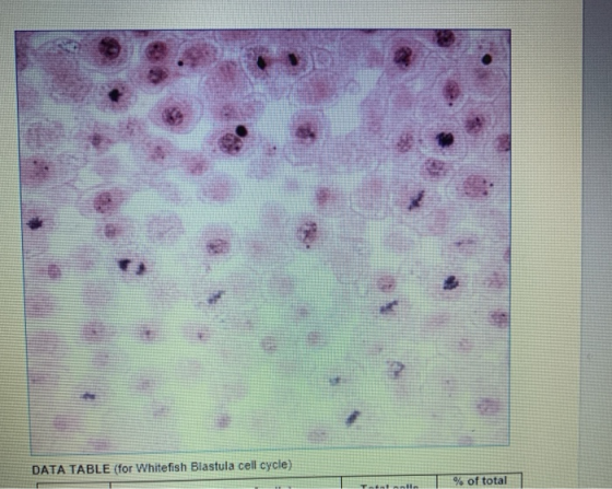
Question 2-  (A) INTERPHASE stage if found most frequently in whitefish blatula
cells.
(A) INTERPHASE stage if found most frequently in whitefish blatula
cells.
Add Answer to:
based on the picture we need to answer these questions
Question 2) Answer the following questions...
BIOS 1705. At-Home Worksheet Cell Division and Cancer (10 points) Name: Below are pictures of cells...
 BIOS 1705. At-Home Worksheet Cell Division and Cancer (10 points) Name: Below are pictures of cells in the various phases of mitosis and the cell cycle from plants for Activity 1 and animal cells for Activity 2. They are in either interphase, prophase, metaphash, anaphase, telophase or cytokinesis. On the line under the picture indicate which phase each cell is in Activity 1 Mitosis in Plant cells -- Onion Root Tip Add-ins Media Links Activity 2 Observing Mitosis in Animal...
Activity 1: Observation of Mitosis in an Onion and a Whitefish Instructions: View the images in...
 Activity 1: Observation of Mitosis in an Onion and a Whitefish Instructions: View the images in Table 1 and Table 2 of the Activity 1 Page to complete blanks below Table 1: Stages of the Cell Cycle in Onion Root Tip Table 2: Stages of Mitosis in Whitefish Blastula *Name Each Stage *Name Each Stage (a) (b) (b) (c) (c) (d) (d) (e) *To name the stages above, choose from: Interphase, Prophase. Metaphase, Anaphase or Telophase/Cytokinesis Identify the number in...
2. In wh o were most of the onion roat tp 7 Based on what you...
 2. In wh o were most of the onion roat tp 7 Based on what you know about all oyole division, what does this imply about the life span of noel? 3. Were there any stages of the cell cycle that you did not observe? How can you explain this using evidence from the cell oyale? 4. As a cell grows, what happens to its surface area to volume ratio? (Hint Think of a balloon being blown up). How does...
Examine the onion root tip slide images on the following pages. There are four images, each...
Examine the onion root tip slide images on the following pages. There are four images, each displaying a different field of view. Pick one of the images, and count the number of cells in each stage. Then count the total number of cells in the image. Record the image you selected and counts in Table 2. Calculate the time spent by a cell in each stage based on the 24 hour cycle: Hours of Stage = 24 x Number of...
5. For this part of the lab you will be observing a whitefish blastula under the...
5. For this part of the lab you will be observing a whitefish blastula under the microscope. A blastula is a developmental stage in many animals. It occurs shortly after fertilization and the formation of the zygote. It is the “ball of cells” stage of development, where cells are dividing rapidly to form the multicellular organism. Note: In this simulation early and late prophase are combined into one stage: prophase. 1. Go to the Virtual Microscope (Links to an external...
Kod of serie Questions Name Aime Guendue Dan Be and we st The D ochis was...
 Kod of serie Questions Name Aime Guendue Dan Be and we st The D ochis was Thoughes to the a uthor you to search Store Tapers loose disput e 3. What is the difference between the terms chromosome and chrom Chromosomes con packed CNA Wire The one hand, Chromatas ONA W S unwound Cow Che a ts function! Spindle,tikers provide a framework & means of attachment that keeps chromosomes organized aligned & assorted during the process of MITOSS 5. During...
the last image is the image for part 2. thank you!! Look through the images of...
 the last image is the image for part 2. thank
you!!
Look through the images of cells below to find examples of cells in the five main stages (interphase, prophase, metaphase, anaphase, telophase). Draw an example of each of these on the sheet to turn in. (Note: I've added a second image of an onion root tip cell in telophase.- notice that a cell wall is starting to form between the two sets of chromosomes.) PART II - TIMING THE...
the last image is the image for part 2. thank
you!!
Look through the images of cells below to find examples of cells in the five main stages (interphase, prophase, metaphase, anaphase, telophase). Draw an example of each of these on the sheet to turn in. (Note: I've added a second image of an onion root tip cell in telophase.- notice that a cell wall is starting to form between the two sets of chromosomes.) PART II - TIMING THE...
2. Match the phase to the description. NOTE: this question might look a little different on...
 2. Match the phase to the description. NOTE: this question might look a little different on the Moodle activity. Read carefully. Phase Hanne Description of Events in the Life of a Cell • Anaphase of muitosis A. The combined phases of GI/Go+S+G2 • Gl phase • G2 phase B. The cell is metabolically active; the cell duplicates argumelles and cytosolie components; the cell size starts to increase the cell makes proteins which will soon be used in DNA synthesis C....
answer this pls asap PART 1: How Long Do Cells Spend in Each stage of Mitosis?...
 answer this pls asap
PART 1: How Long Do Cells Spend in Each stage of Mitosis? In this activity, you will be presented with cells from the tip of an onion root. You will classify each cell based on what phase it is in. At the end you will count up the cells found in each phase and use those numbers to predict how much time a dividing cell spends in each phase. You can base your calculation on a...
answer this pls asap
PART 1: How Long Do Cells Spend in Each stage of Mitosis? In this activity, you will be presented with cells from the tip of an onion root. You will classify each cell based on what phase it is in. At the end you will count up the cells found in each phase and use those numbers to predict how much time a dividing cell spends in each phase. You can base your calculation on a...
The vesicles that form between the two plant daughter cells are small buttles of phospholipid membrane...
 The vesicles that form between the two plant daughter cells
are small buttles of phospholipid membrane formed by the Golgi
apparatus . When these vesicles fuse , what do they become? HINT:
The bubbles are made of phospholipids.
And please fill the table in the picture.
Procedure 8.5 ESTIMATE THE TIME ELAPSED DURING THE PHASES OF MITOSIS Do all phases of the cell cycle require the same amount of time? The answer can be found by counting the number of...
The vesicles that form between the two plant daughter cells
are small buttles of phospholipid membrane formed by the Golgi
apparatus . When these vesicles fuse , what do they become? HINT:
The bubbles are made of phospholipids.
And please fill the table in the picture.
Procedure 8.5 ESTIMATE THE TIME ELAPSED DURING THE PHASES OF MITOSIS Do all phases of the cell cycle require the same amount of time? The answer can be found by counting the number of...
 BIOS 1705. At-Home Worksheet Cell Division and Cancer (10 points) Name: Below are pictures of cells in the various phases of mitosis and the cell cycle from plants for Activity 1 and animal cells for Activity 2. They are in either interphase, prophase, metaphash, anaphase, telophase or cytokinesis. On the line under the picture indicate which phase each cell is in Activity 1 Mitosis in Plant cells -- Onion Root Tip Add-ins Media Links Activity 2 Observing Mitosis in Animal...
 Activity 1: Observation of Mitosis in an Onion and a Whitefish Instructions: View the images in Table 1 and Table 2 of the Activity 1 Page to complete blanks below Table 1: Stages of the Cell Cycle in Onion Root Tip Table 2: Stages of Mitosis in Whitefish Blastula *Name Each Stage *Name Each Stage (a) (b) (b) (c) (c) (d) (d) (e) *To name the stages above, choose from: Interphase, Prophase. Metaphase, Anaphase or Telophase/Cytokinesis Identify the number in...
 2. In wh o were most of the onion roat tp 7 Based on what you know about all oyole division, what does this imply about the life span of noel? 3. Were there any stages of the cell cycle that you did not observe? How can you explain this using evidence from the cell oyale? 4. As a cell grows, what happens to its surface area to volume ratio? (Hint Think of a balloon being blown up). How does...
 Kod of serie Questions Name Aime Guendue Dan Be and we st The D ochis was Thoughes to the a uthor you to search Store Tapers loose disput e 3. What is the difference between the terms chromosome and chrom Chromosomes con packed CNA Wire The one hand, Chromatas ONA W S unwound Cow Che a ts function! Spindle,tikers provide a framework & means of attachment that keeps chromosomes organized aligned & assorted during the process of MITOSS 5. During...
 the last image is the image for part 2. thank
you!!
Look through the images of cells below to find examples of cells in the five main stages (interphase, prophase, metaphase, anaphase, telophase). Draw an example of each of these on the sheet to turn in. (Note: I've added a second image of an onion root tip cell in telophase.- notice that a cell wall is starting to form between the two sets of chromosomes.) PART II - TIMING THE...
the last image is the image for part 2. thank
you!!
Look through the images of cells below to find examples of cells in the five main stages (interphase, prophase, metaphase, anaphase, telophase). Draw an example of each of these on the sheet to turn in. (Note: I've added a second image of an onion root tip cell in telophase.- notice that a cell wall is starting to form between the two sets of chromosomes.) PART II - TIMING THE...
 2. Match the phase to the description. NOTE: this question might look a little different on the Moodle activity. Read carefully. Phase Hanne Description of Events in the Life of a Cell • Anaphase of muitosis A. The combined phases of GI/Go+S+G2 • Gl phase • G2 phase B. The cell is metabolically active; the cell duplicates argumelles and cytosolie components; the cell size starts to increase the cell makes proteins which will soon be used in DNA synthesis C....
 answer this pls asap
PART 1: How Long Do Cells Spend in Each stage of Mitosis? In this activity, you will be presented with cells from the tip of an onion root. You will classify each cell based on what phase it is in. At the end you will count up the cells found in each phase and use those numbers to predict how much time a dividing cell spends in each phase. You can base your calculation on a...
answer this pls asap
PART 1: How Long Do Cells Spend in Each stage of Mitosis? In this activity, you will be presented with cells from the tip of an onion root. You will classify each cell based on what phase it is in. At the end you will count up the cells found in each phase and use those numbers to predict how much time a dividing cell spends in each phase. You can base your calculation on a...
 The vesicles that form between the two plant daughter cells
are small buttles of phospholipid membrane formed by the Golgi
apparatus . When these vesicles fuse , what do they become? HINT:
The bubbles are made of phospholipids.
And please fill the table in the picture.
Procedure 8.5 ESTIMATE THE TIME ELAPSED DURING THE PHASES OF MITOSIS Do all phases of the cell cycle require the same amount of time? The answer can be found by counting the number of...
The vesicles that form between the two plant daughter cells
are small buttles of phospholipid membrane formed by the Golgi
apparatus . When these vesicles fuse , what do they become? HINT:
The bubbles are made of phospholipids.
And please fill the table in the picture.
Procedure 8.5 ESTIMATE THE TIME ELAPSED DURING THE PHASES OF MITOSIS Do all phases of the cell cycle require the same amount of time? The answer can be found by counting the number of...
Most questions answered within 3 hours.
-
You are attempting to calculate a firm’s free cash flow to
equity. You know the following...
asked 33 minutes ago -
the following reaction occurs in a balloon containing
N2O2 gas
N2O4(g)=2NO2(g)
will the volume of the...
asked 1 hour ago -
answer the questions throughout this program
public class Day implements Comparable {
Private Boolean atWork;...
asked 1 hour ago -
This is C++ code for parking fee management program
#include <iostream>
#include <iomanip>
using namespace std;...
asked 1 hour ago -
The free energy change for the following reaction at 25 °C, when
[Sn2+] = 1.17 M...
asked 3 hours ago -
An MNE is this kind of industry when competition in one country
is essentially independent of...
asked 4 hours ago -
. For this set of questions, determine what
proportion of a normal distribution is located betweeneach...
asked 5 hours ago -
A college student is employed as a door-to-door newspaper
salesman. Historical data suggests that the student...
asked 6 hours ago -
MATLAB HW 11 problem using Switch Case and Input commands
Write a script file that calculates...
asked 5 hours ago -
Considering gravitational time dilation, calculate the time that
passes in Earth’s surface while 1 hour passes...
asked 6 hours ago -
Minitab Problem: Take the Lake Hume June rainfall data and find
use the processes outlined in...
asked 7 hours ago -
X Company is trying to decide whether to continue using old
equipment to make Product A...
asked 7 hours ago