
Homework Answers
d) Propane CH3-CH2-CH3
As the spin-spin coupling is absent, the peaks will not split.
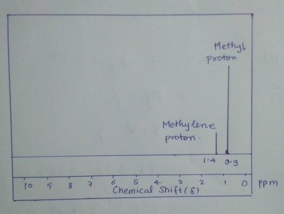
The difference in the peak lengths is due to:
Methyl group CH3 has three hydrogen atoms connected to one carbon atom and there are two methyl groups present is propane.
Methylene group CH2 has two hydrogen atoms connected to one carbon atom and there is only one methylene group present in propane.

# Peak area ratio
Peak area of Methyl proton / Peak area of Methylene proton = (Length of peak x width of peak)methyl / (Length of peak x width of peak)methylene
Add Answer to:
(4 pts) Factoring in the spin-spin coupling, draw the expected NMR spectrum of the methyl protons...
Shown below is the peak list of chemical shifts, multiplicities, numbers of protons (from integration), and...
 Shown below is the peak list of chemical shifts, multiplicities,
numbers of protons (from integration), and coupling constants for
the 1H NMR spectrum in Question 4.
a) Draw the chemical structure of trans-4-nitrochalcone. Circle
the proton or protons that you would attempt to identify in the 1H
NMR spectrum as evidence that the expected trans-isomer was formed
(rather than the cis-isomer).
b) Identify the peak or peaks in the peak list that
correspond(s) to the proton or protons circled in...
Shown below is the peak list of chemical shifts, multiplicities,
numbers of protons (from integration), and coupling constants for
the 1H NMR spectrum in Question 4.
a) Draw the chemical structure of trans-4-nitrochalcone. Circle
the proton or protons that you would attempt to identify in the 1H
NMR spectrum as evidence that the expected trans-isomer was formed
(rather than the cis-isomer).
b) Identify the peak or peaks in the peak list that
correspond(s) to the proton or protons circled in...
Draw the structure of the compound C6H5ClO from its proton (1H) NMR spectrum below. First-order spin-spin...
 Draw the structure of the compound
C6H5ClO from its proton
(1H) NMR spectrum below.
First-order spin-spin splitting rules and equal coupling
constants can be assumed. (Detailed analysis of any non-first order
portions of the spectrum will not be required.)
Integral ratios to the nearest whole number are (left to right)
2:2:1.
Flash Installation and Troubleshooting
graph data : 7.18 2153HZ 6.77. 2031Hz 5.25 1575HZ .01. 2 Hz
Draw the structure of the compound CH CIO from its proton ("H) NMR...
Draw the structure of the compound
C6H5ClO from its proton
(1H) NMR spectrum below.
First-order spin-spin splitting rules and equal coupling
constants can be assumed. (Detailed analysis of any non-first order
portions of the spectrum will not be required.)
Integral ratios to the nearest whole number are (left to right)
2:2:1.
Flash Installation and Troubleshooting
graph data : 7.18 2153HZ 6.77. 2031Hz 5.25 1575HZ .01. 2 Hz
Draw the structure of the compound CH CIO from its proton ("H) NMR...
H NMR Spectrum: For each signal: 1.) Identify its environment 2.) Identify its spin-spin coupling...
 H NMR Spectrum:
For each signal:
1.) Identify its environment
2.) Identify its spin-spin coupling (identify how many protons
are 3 bonds away, causing the coupling)
3.) Identify its integration value
Why is the peak at 6.3 ppm broadened?
Explain why there are 2 doublets in the aromatic region, but 4
aromatic protons on benzocaine
Why is the quartet at ~4.3 ppm so far downfield compared to the
triplet at ~ 1.3 ppm?
What is the purpose of using sulfuric...
H NMR Spectrum:
For each signal:
1.) Identify its environment
2.) Identify its spin-spin coupling (identify how many protons
are 3 bonds away, causing the coupling)
3.) Identify its integration value
Why is the peak at 6.3 ppm broadened?
Explain why there are 2 doublets in the aromatic region, but 4
aromatic protons on benzocaine
Why is the quartet at ~4.3 ppm so far downfield compared to the
triplet at ~ 1.3 ppm?
What is the purpose of using sulfuric...
Predicting the Spectrum MR and C NMR) the expected 'H NMR number of si spectrum for...
 Predicting the Spectrum MR and C NMR) the expected 'H NMR number of si spectrum for the following molecule. Be sure to include appropriate for the following molecule. Be sure to er of signals, show spisina ar try to indicate relative Integration by arca un (label peak with some splitting and try to indicate relative Integration by area under peak peak with correct number for clarity in tabel all the unique hydrogens with letters and assign them to the peaks...
Draw the structure of the compound C3H4Cl2O from its proton NMR spectrum below. Please explain. Draw...
 Draw the structure of the compound C3H4Cl2O from its proton NMR
spectrum below. Please explain.
Draw the structure of the compound CH CI O from its proton (1) NMR spectrum below. Fust-order spin-spin splitting rules and equal coupling constants can be assumed. (Detailed analysis of any non-first order portions of the spectrum will not be required) Integral ratios to the nearest whole number are left to right) 1:3 Flash Installation and Troubleshooting Used with from At Checa Co Inc (purty...
Draw the structure of the compound C3H4Cl2O from its proton NMR
spectrum below. Please explain.
Draw the structure of the compound CH CI O from its proton (1) NMR spectrum below. Fust-order spin-spin splitting rules and equal coupling constants can be assumed. (Detailed analysis of any non-first order portions of the spectrum will not be required) Integral ratios to the nearest whole number are left to right) 1:3 Flash Installation and Troubleshooting Used with from At Checa Co Inc (purty...
Draw the structure of the compound C3H3CI3 from its proton (H) NMR spectrum below. First-order spin-spin...
 Draw the structure of the compound C3H3CI3 from its proton (H) NMR spectrum below. First-order spin-spin splitting rules and equal coupling constants can be assumed. (Detailed analysis of any non-first order portions of the spectrum will not be required.) Integral ratios to the nearest whole number are (left to right) 1:2 Flash Installation and Troubleshooting Solvent CDCl Used with permission from Aldrich Chemical Co., Inc (Impurities at 3.34, 4.05, & 4.07 ppm) Chemical shift x5 Y ZOOM MEASURE 15 0.0...
The proton NMR spectrum for a compound with the formula C,H,O, is shown below along with...
 The proton NMR spectrum for a compound with the formula C,H,O, is shown below along with the spectral data in tabular form. (It may be necessary to expand (zoom) some of the 'H signals to view spin-spin splitting details.) This compound exhibits strong infrared absorption at 1727 and 1195 em plus a medium intensity band at 1637 cm! 13C NMR Data 'H NMR Data Proton Shift Relative Area Normal Carbon DEPT-135 DEPT-90 14.2 Positive No peak 60.4 Negative No peak...
(References The proton NMR spectrum for a compound with the formula C,H,O is shown below along...
 (References The proton NMR spectrum for a compound with the formula C,H,O is shown below along with the spectral data in tabular form. (It may be necessary to expand (zoom) some of the 'H signals to view spin-spin splitting details.) Intensity 258 $ $ 488 14 13 12 11 10 9 8 7 6 5 4 3 Chemical Shift (oppm 2 1 0 . Reproduced with permission from Sim. Aldri Ca LLC The IH signal at 2.15 ppm is an...
Draw the expected H NMR spectrum for each molecule. Pay attention to the chemical shift and...
 Draw the expected H NMR spectrum for each molecule. Pay
attention to the chemical shift and splitting. Mark the integration
of each peak. Then assign each peak in the NMR to hydrogen atoms in
the structure.
HyC 0 ppm 192 t2 .3 +5 o t6 O=C +7 C=O +8
Draw the expected H NMR spectrum for each molecule. Pay
attention to the chemical shift and splitting. Mark the integration
of each peak. Then assign each peak in the NMR to hydrogen atoms in
the structure.
HyC 0 ppm 192 t2 .3 +5 o t6 O=C +7 C=O +8
Create a table with the headings Proton Label, Theoretical Chemical Shift (8 ppm), multipilicity, coupling, &...
 Create a table with the headings Proton Label,
Theoretical Chemical Shift (8 ppm), multipilicity, coupling, &
theoretical J value (Hz) for Acetylsalicylic acid using the labels
shown on the structure below. On the spectrum you made during the
lab, assign the protons by labeling the peaks on the spectrum with
the correct proton label.
3. Create a table with the headings Proton Label, Theoretical Chemical Shift (8 ppm), multipilicity, coupling, & theoretical J value (Hz) for Acetylsalicylic acid using the...
Create a table with the headings Proton Label,
Theoretical Chemical Shift (8 ppm), multipilicity, coupling, &
theoretical J value (Hz) for Acetylsalicylic acid using the labels
shown on the structure below. On the spectrum you made during the
lab, assign the protons by labeling the peaks on the spectrum with
the correct proton label.
3. Create a table with the headings Proton Label, Theoretical Chemical Shift (8 ppm), multipilicity, coupling, & theoretical J value (Hz) for Acetylsalicylic acid using the...
 Shown below is the peak list of chemical shifts, multiplicities,
numbers of protons (from integration), and coupling constants for
the 1H NMR spectrum in Question 4.
a) Draw the chemical structure of trans-4-nitrochalcone. Circle
the proton or protons that you would attempt to identify in the 1H
NMR spectrum as evidence that the expected trans-isomer was formed
(rather than the cis-isomer).
b) Identify the peak or peaks in the peak list that
correspond(s) to the proton or protons circled in...
Shown below is the peak list of chemical shifts, multiplicities,
numbers of protons (from integration), and coupling constants for
the 1H NMR spectrum in Question 4.
a) Draw the chemical structure of trans-4-nitrochalcone. Circle
the proton or protons that you would attempt to identify in the 1H
NMR spectrum as evidence that the expected trans-isomer was formed
(rather than the cis-isomer).
b) Identify the peak or peaks in the peak list that
correspond(s) to the proton or protons circled in...
 Draw the structure of the compound
C6H5ClO from its proton
(1H) NMR spectrum below.
First-order spin-spin splitting rules and equal coupling
constants can be assumed. (Detailed analysis of any non-first order
portions of the spectrum will not be required.)
Integral ratios to the nearest whole number are (left to right)
2:2:1.
Flash Installation and Troubleshooting
graph data : 7.18 2153HZ 6.77. 2031Hz 5.25 1575HZ .01. 2 Hz
Draw the structure of the compound CH CIO from its proton ("H) NMR...
Draw the structure of the compound
C6H5ClO from its proton
(1H) NMR spectrum below.
First-order spin-spin splitting rules and equal coupling
constants can be assumed. (Detailed analysis of any non-first order
portions of the spectrum will not be required.)
Integral ratios to the nearest whole number are (left to right)
2:2:1.
Flash Installation and Troubleshooting
graph data : 7.18 2153HZ 6.77. 2031Hz 5.25 1575HZ .01. 2 Hz
Draw the structure of the compound CH CIO from its proton ("H) NMR...
 H NMR Spectrum:
For each signal:
1.) Identify its environment
2.) Identify its spin-spin coupling (identify how many protons
are 3 bonds away, causing the coupling)
3.) Identify its integration value
Why is the peak at 6.3 ppm broadened?
Explain why there are 2 doublets in the aromatic region, but 4
aromatic protons on benzocaine
Why is the quartet at ~4.3 ppm so far downfield compared to the
triplet at ~ 1.3 ppm?
What is the purpose of using sulfuric...
H NMR Spectrum:
For each signal:
1.) Identify its environment
2.) Identify its spin-spin coupling (identify how many protons
are 3 bonds away, causing the coupling)
3.) Identify its integration value
Why is the peak at 6.3 ppm broadened?
Explain why there are 2 doublets in the aromatic region, but 4
aromatic protons on benzocaine
Why is the quartet at ~4.3 ppm so far downfield compared to the
triplet at ~ 1.3 ppm?
What is the purpose of using sulfuric...
 Predicting the Spectrum MR and C NMR) the expected 'H NMR number of si spectrum for the following molecule. Be sure to include appropriate for the following molecule. Be sure to er of signals, show spisina ar try to indicate relative Integration by arca un (label peak with some splitting and try to indicate relative Integration by area under peak peak with correct number for clarity in tabel all the unique hydrogens with letters and assign them to the peaks...
 Draw the structure of the compound C3H4Cl2O from its proton NMR
spectrum below. Please explain.
Draw the structure of the compound CH CI O from its proton (1) NMR spectrum below. Fust-order spin-spin splitting rules and equal coupling constants can be assumed. (Detailed analysis of any non-first order portions of the spectrum will not be required) Integral ratios to the nearest whole number are left to right) 1:3 Flash Installation and Troubleshooting Used with from At Checa Co Inc (purty...
Draw the structure of the compound C3H4Cl2O from its proton NMR
spectrum below. Please explain.
Draw the structure of the compound CH CI O from its proton (1) NMR spectrum below. Fust-order spin-spin splitting rules and equal coupling constants can be assumed. (Detailed analysis of any non-first order portions of the spectrum will not be required) Integral ratios to the nearest whole number are left to right) 1:3 Flash Installation and Troubleshooting Used with from At Checa Co Inc (purty...
 Draw the structure of the compound C3H3CI3 from its proton (H) NMR spectrum below. First-order spin-spin splitting rules and equal coupling constants can be assumed. (Detailed analysis of any non-first order portions of the spectrum will not be required.) Integral ratios to the nearest whole number are (left to right) 1:2 Flash Installation and Troubleshooting Solvent CDCl Used with permission from Aldrich Chemical Co., Inc (Impurities at 3.34, 4.05, & 4.07 ppm) Chemical shift x5 Y ZOOM MEASURE 15 0.0...
 The proton NMR spectrum for a compound with the formula C,H,O, is shown below along with the spectral data in tabular form. (It may be necessary to expand (zoom) some of the 'H signals to view spin-spin splitting details.) This compound exhibits strong infrared absorption at 1727 and 1195 em plus a medium intensity band at 1637 cm! 13C NMR Data 'H NMR Data Proton Shift Relative Area Normal Carbon DEPT-135 DEPT-90 14.2 Positive No peak 60.4 Negative No peak...
 (References The proton NMR spectrum for a compound with the formula C,H,O is shown below along with the spectral data in tabular form. (It may be necessary to expand (zoom) some of the 'H signals to view spin-spin splitting details.) Intensity 258 $ $ 488 14 13 12 11 10 9 8 7 6 5 4 3 Chemical Shift (oppm 2 1 0 . Reproduced with permission from Sim. Aldri Ca LLC The IH signal at 2.15 ppm is an...
 Draw the expected H NMR spectrum for each molecule. Pay
attention to the chemical shift and splitting. Mark the integration
of each peak. Then assign each peak in the NMR to hydrogen atoms in
the structure.
HyC 0 ppm 192 t2 .3 +5 o t6 O=C +7 C=O +8
Draw the expected H NMR spectrum for each molecule. Pay
attention to the chemical shift and splitting. Mark the integration
of each peak. Then assign each peak in the NMR to hydrogen atoms in
the structure.
HyC 0 ppm 192 t2 .3 +5 o t6 O=C +7 C=O +8
 Create a table with the headings Proton Label,
Theoretical Chemical Shift (8 ppm), multipilicity, coupling, &
theoretical J value (Hz) for Acetylsalicylic acid using the labels
shown on the structure below. On the spectrum you made during the
lab, assign the protons by labeling the peaks on the spectrum with
the correct proton label.
3. Create a table with the headings Proton Label, Theoretical Chemical Shift (8 ppm), multipilicity, coupling, & theoretical J value (Hz) for Acetylsalicylic acid using the...
Create a table with the headings Proton Label,
Theoretical Chemical Shift (8 ppm), multipilicity, coupling, &
theoretical J value (Hz) for Acetylsalicylic acid using the labels
shown on the structure below. On the spectrum you made during the
lab, assign the protons by labeling the peaks on the spectrum with
the correct proton label.
3. Create a table with the headings Proton Label, Theoretical Chemical Shift (8 ppm), multipilicity, coupling, & theoretical J value (Hz) for Acetylsalicylic acid using the...
Most questions answered within 3 hours.
-
1.With time, an appreciation in the value of the nation's
currency in the foreign exchange market...
asked 30 seconds ago -
Personal selling is of special importance in B2B marketing. What
has been the impact of the...
asked 4 minutes ago -
In 2003, an organization surveyed 1 comma 510 adult Americans
and asked about a certain war,...
asked 11 minutes ago -
Blue colorin fireworks is often achieved by heating copper(I)
chloride (CuCl) to about 1200°C. Then the...
asked 6 minutes ago -
Coperto Insurers of Davenport, Iowa offers four different
insurance plans, all of which are equally popular...
asked 14 minutes ago -
A 50 mL graduated cylinder contains 25.0 mL of water. A 42.5040
g piece of gold...
asked 15 minutes ago -
Leo’s Lamp Store sells table and floor lamps. For a floor lamp,
the holding cost is...
asked 17 minutes ago -
Mark Gershon, owner of a musical instrument distributorship,
thinks that demand for guitars may be related...
asked 29 minutes ago -
Suppose that you were asked to construct a 95% confidence
interval based on the standard normal...
asked 36 minutes ago -
3 - What decimal number does the bit pattern 11001100 represent
if it is a:
•...
asked 38 minutes ago -
The copper(II) ion is acidic whereas the acetate ion is basic.
However, copper acetate is acidic....
asked 34 minutes ago -
A 48.53 mL volume of 1.00 M HCl was mixed with 47.70 mL of 2.00
M...
asked 56 minutes ago