Directions: There are two parts for this lab. The first part is working with the Microscope slides. The second part is working with media.
Task 1 – Bacteriology Survey Microscope Slides
Directions: There are eight different bacterium you need to
research.
Below is the following bacterium you need to describe and include
an image for.
• Streptococcus
• Treponema
• Neisseria gonorrhoeae
• Klebisella pneumonia
• Unstained Bacillus
• Bacillus cereus
• Clostridium tetani
• Escherichia coli
Task 2 – Observing Media in a Petri Dish
Directions: Below are examples of media that has been plated and
share descriptions of each. Please answer the questions under each
image description.
Observing bacteria in a petri dish
Students should examine cultures in containers, which have been
taped and closed. Colony morphology is a method that scientists use
to describe the characteristics of an individual colony of bacteria
growing on agar in a Petri dish. It can be used to help to identify
them.
Different types of bacteria will produce different-looking
colonies, some colonies may be colored, some colonies are circular
in shape, and others are irregular. A specific terminology is used
to describe common colony types. These are:
• Form - What is the basic shape of the colony? For
example, circular, filamentous, etc.
• Size – The diameter of the colony. Tiny colonies are
referred to as punctiform.
• Elevation - This describes the side view of a colony.
Turn the Petri dish on end.
• Margin/border – The edge of a colony. What is the
magnified shape of the edge of the colony?
• Surface - How does the surface of the colony appear?
For example, smooth, glistening, rough, wrinkled or dull.
• Opacity - For example, transparent (clear), opaque,
translucent (like looking through frosted glass), etc.
• Colour - (pigmentation) - For example, white, buff,
red, purple, etc.
Each distinct colony represents an individual bacterial cell or
group that has divided repeatedly. Being kept in one place, the
resulting cells have accumulated to form a visible patch. Most
bacterial colonies appear white or a creamy yellow in colour, and
are fairly circular in shape.


A swab from a bin spread directly onto nutrient agar.
Colonies differ in their shape, size, colour and texture. Can you
count how many different colony types there are? Use the diagrams
on colony morphology to help you interpret your plate.
idophores
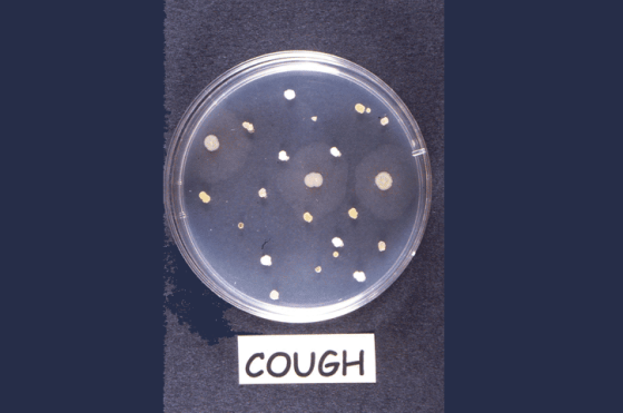
A cough that was aimed directly onto nutrient agar.
Colonies differ in their shape, size, colour and texture. Can you
count how many different colony types there are? Use the diagrams
on colony morphology to help you interpret your plate.
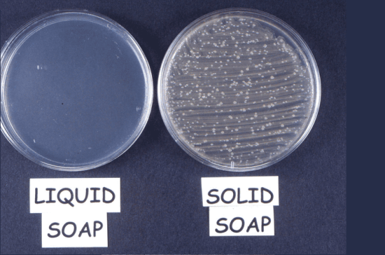
A sample of liquid soap spread onto nutrient agar
A sample of liquid soap spread onto nutrient agar and a swab from a
bar of solid soap also spread onto nutrient agar. Soaps are not
designed to kill microbes. They help to wash them of your skin,
better than water alone. Why do you think that the solid soap (kept
by the sink and handled regularly) had more bacteria living on it
than the liquid soap (kept in a dispenser, so not handled) which
had none? You may be interested to know that while soaps do not
kill microbes they can be quite a good medium for them to grow
on!

A streak plate to isolate single colonies of a specific
bacterium
A streak plate to isolate single colonies of a specific bacterium
found living on a sample of paper. Paper is not a good object for
bacteria to live on as it cannot sustain their growth. The bacteria
found on the paper are known as transient i.e. they are just
passing the time until a better place to live comes along. Handling
the paper would transfer the resident bacteria from a person's hand
to the paper.
Based on the examples provided, which stood out to your the most or
caught your attention I should say? How do these images play an
important part in understanding microbes within
microbiology?
Homework Answers
Task 1
1. Streptococcus is a genus of Gram-positive coccus or spherical bacteria that belongs to the family Streptococcaceae, within the order Lactobacillales, in the phylum Firmicutes. Cell division in streptococci occurs along a single axis, so as they grow, they tend to form pairs or chains that may appear bent or twisted.
 2. Treponema is a genus of
spiral-shaped bacteria. The major treponeme species of human
pathogens is Treponema pallidum, whose subspecies are responsible
for diseases such as syphilis, bejel, and yaws. Treponema carateum
is the cause of pinta.
2. Treponema is a genus of
spiral-shaped bacteria. The major treponeme species of human
pathogens is Treponema pallidum, whose subspecies are responsible
for diseases such as syphilis, bejel, and yaws. Treponema carateum
is the cause of pinta.
3. Neisseria gonorrhoeae, also known as gonococcus (singular),
or gonococci (plural) is a species of Gram-negative diplococci
bacteria isolated by Albert Neisser in 1879
4. Klebsiella pneumoniae is a Gram-negative, non-motile, encapsulated, lactose-fermenting, facultative anaerobic, rod-shaped bacterium. It appears as a mucoid lactose fermenter on MacConkey agar.

5. Bacillus (Latin "stick") is a genus of Gram-positive,
rod-shaped bacteria, a member of the phylum Firmicutes, with 266
named species.  6. Bacillus cereus is a
Gram-positive, rod-shaped, facultatively anaerobic, motile,
beta-hemolytic, spore forming bacterium commonly found in soil and
food. The specific name, cereus, meaning "waxy" in Latin, refers to
the appearance of colonies grown on blood agar.
6. Bacillus cereus is a
Gram-positive, rod-shaped, facultatively anaerobic, motile,
beta-hemolytic, spore forming bacterium commonly found in soil and
food. The specific name, cereus, meaning "waxy" in Latin, refers to
the appearance of colonies grown on blood agar. 7. Clostridium tetani is a
common soil bacterium and the causative agent of tetanus. When
growing in soil, C. tetani is rod-shaped and up to 2.5 μm long.
However, when forming spores, C. tetani becomes substantially
enlarged at one end, resembling a tennis racket or
drumstick.
7. Clostridium tetani is a
common soil bacterium and the causative agent of tetanus. When
growing in soil, C. tetani is rod-shaped and up to 2.5 μm long.
However, when forming spores, C. tetani becomes substantially
enlarged at one end, resembling a tennis racket or
drumstick. 8. coli (Escherichia coli), is
a type of bacteria that normally lives in your intestines. It's
also found in the gut of some animals. Most types of E. coli are
harmless and even help keep your digestive tract healthy. But some
strains can cause diarrhea if you eat contaminated food or drink
fouled water.
8. coli (Escherichia coli), is
a type of bacteria that normally lives in your intestines. It's
also found in the gut of some animals. Most types of E. coli are
harmless and even help keep your digestive tract healthy. But some
strains can cause diarrhea if you eat contaminated food or drink
fouled water.
Add Answer to:
Directions: There are two parts for this lab. The first part is
working with the Microscope...
Micrebes in the Environment QUESTIONS What in the advantage of using Petri plate rather thon tet...
 Micrebes in the Environment QUESTIONS What in the advantage of using Petri plate rather thon tet t in mieriogy? 2What are bacteria uaing for nutrienta in satrient agsr What is the perpoe of the agar? CRITICAL THINKING Did all the organiems living in or on the environmente aompled gw on your nutrient agar Briefly explain. 1. 2 How could you determine whether the turbidity in your nutrient broth tube was from a mixture of diferent microbes or from the growth...
Lab Report-Carbohydrates 1. Purpose 2. Special Media for Isolating Bacteria (Lab #12) a. Why are dyes...
 Lab Report-Carbohydrates 1. Purpose 2. Special Media for Isolating Bacteria (Lab #12) a. Why are dyes such as phenol red, eosin or methylene blue added to the media? b. How does the bacterium change the media (i.e color of agar or colonies) after incubation? C. In this experiment, which media are selective, and which are differential? d. How did the results observe on the mannitol salt agar and EMB agar correlate to the Gram reaction of the bacteria? e. What...
GROUP (Report 1 Growth Curve 1) The following table shows the measurements of bacterial growth in...
 GROUP (Report 1 Growth Curve 1) The following table shows the measurements of bacterial growth in a specific period by the color Spectrophotometer Time time 3 hour Growth measurement 7.56 7.56 30.25 32.25 6 hour 9 hour 12 hour 32.25 15 hour 2.69 A) Draw the growth curve for bacteria B) Write a discussion of the results (Abbreviator) 2) The plate was inoculation with a fungal disc 1 cm, and then incubated for 7 days and 25C * The following...
I'm not sure if I completed question 3 correctly. Also, I need help determing question 4....
 I'm
not sure if I completed question 3 correctly. Also, I need help
determing question 4.
Y-0.301 6.51-hardts.al 3. A solution containing 100 mL of bacterial broth is subcultured onto 5 plates using the pour-plate method. Each plate receives broth according to the following Table. The Table also lists the results colony counts after 24 hours of incubation. Calculate the number of bacteria per milliliter of the bacterial broth for each of the dilutions. Plate Volume Broth Colonies 331 2...
I'm
not sure if I completed question 3 correctly. Also, I need help
determing question 4.
Y-0.301 6.51-hardts.al 3. A solution containing 100 mL of bacterial broth is subcultured onto 5 plates using the pour-plate method. Each plate receives broth according to the following Table. The Table also lists the results colony counts after 24 hours of incubation. Calculate the number of bacteria per milliliter of the bacterial broth for each of the dilutions. Plate Volume Broth Colonies 331 2...
infectious disease lab summary protocol. Questions that need to be answered are attached, aswell as the...
 infectious disease lab summary protocol. Questions that need to be
answered are attached, aswell as the lab manuel
Name: ID: Protocol summary for: (Maximum 2 pages, Total = 10 marks) 1) Describe, in a sentence or two, the main objective(s) of this lab. (1) 2) What techniques will you be using in this lab? (2) 3) How many microorganisms will you be working with in this lab? (1) 4) Over the course of this lab (1", 24, ... week), which...
infectious disease lab summary protocol. Questions that need to be
answered are attached, aswell as the lab manuel
Name: ID: Protocol summary for: (Maximum 2 pages, Total = 10 marks) 1) Describe, in a sentence or two, the main objective(s) of this lab. (1) 2) What techniques will you be using in this lab? (2) 3) How many microorganisms will you be working with in this lab? (1) 4) Over the course of this lab (1", 24, ... week), which...
Multiple Choice. Highlight the single correct answer choice. 1. Streak plates are useful in microbiology to...
Multiple Choice. Highlight the single correct answer choice. 1. Streak plates are useful in microbiology to __________. quantify the number of bacteria measure turbidity identify bacteria determine cell shape 2. In the streak-plate technique, the intent is to isolate bacteria by dilution in theory by __________. dilution on a solid surface separating cells within the solid surface using a pipette dilution in water blanks 3. A pure culture consists of which of the following? one genus of microbe one species...
please help me with letter B. Value Ulla ination of Living EXERCISE 3: MICROE b. Holding...
 please help me with letter B.
Value Ulla ination of Living EXERCISE 3: MICROE b. Holding the flask at an angle, remove the stop- per with the fourth and fifth fingers of your oth- er hand. Heat the mouth of the flask by passing it through the flame three times (FIGURE 2a). Why is it necessary to keep the flask at an an- gle through this procedure? (a) Remove theflask. c. Remove the cover from the first dish with the...
please help me with letter B.
Value Ulla ination of Living EXERCISE 3: MICROE b. Holding the flask at an angle, remove the stop- per with the fourth and fifth fingers of your oth- er hand. Heat the mouth of the flask by passing it through the flame three times (FIGURE 2a). Why is it necessary to keep the flask at an an- gle through this procedure? (a) Remove theflask. c. Remove the cover from the first dish with the...
Lab Questions at End (picture 3) QUESTIONS This week in lab you will learn an important...
 Lab
Questions at End (picture 3)
QUESTIONS
This week in lab you will learn an important technique that microbiologists use to isolate one bacterial species from the population of other microbes it was living with in the natural environment. In most environments there are mixed populations of bacteria. For instance, 600 different types of bacteria have been isolated and identified in the mouths of humans. In soil or water there are also many types of bacteria present. In order to...
Lab
Questions at End (picture 3)
QUESTIONS
This week in lab you will learn an important technique that microbiologists use to isolate one bacterial species from the population of other microbes it was living with in the natural environment. In most environments there are mixed populations of bacteria. For instance, 600 different types of bacteria have been isolated and identified in the mouths of humans. In soil or water there are also many types of bacteria present. In order to...
e) Can you identify any wildtype (nonmutant) colonies in the figures? Explain your answer. f) Can...
 e) Can you identify any wildtype (nonmutant) colonies in the figures? Explain your answer. f) Can you identify any standard (non-conditional) mutants in the figures? Explain your answer. g) Can you identify any conditional mutants in the figures? Explain your answer. 23°C mutant colony in which cells proliferate at the cooler, permissive temperature but fail to proliferate at the warmer, nonpermissive temperature 23°C colonies replicated onto two identical plates and incubated at two different temperatures mutagenized cells plated out in...
Use the Broch Care Putter (fig. 4) chart to fill in the datatable Table 3. Hroth...
 Use the Broch Care Putter (fig. 4) chart to fill in the datatable Table 3. Hroth Liquid Culture Growth Patterns (Y/N) Environmental Samples Lab Hedy Sampler Belybutton Sink Turbidity Results: Use the Colony Description (fig. 3) chart to fill in the data tables. Use a new line for each different colony type found on the environmental and body part samples. Table 1: Environmental sample Colony Descriptions Whole colony Margin Elevation Pigment appearance Area sampled Lab sink HH 217 Incubated at...
 Micrebes in the Environment QUESTIONS What in the advantage of using Petri plate rather thon tet t in mieriogy? 2What are bacteria uaing for nutrienta in satrient agsr What is the perpoe of the agar? CRITICAL THINKING Did all the organiems living in or on the environmente aompled gw on your nutrient agar Briefly explain. 1. 2 How could you determine whether the turbidity in your nutrient broth tube was from a mixture of diferent microbes or from the growth...
 Lab Report-Carbohydrates 1. Purpose 2. Special Media for Isolating Bacteria (Lab #12) a. Why are dyes such as phenol red, eosin or methylene blue added to the media? b. How does the bacterium change the media (i.e color of agar or colonies) after incubation? C. In this experiment, which media are selective, and which are differential? d. How did the results observe on the mannitol salt agar and EMB agar correlate to the Gram reaction of the bacteria? e. What...
 GROUP (Report 1 Growth Curve 1) The following table shows the measurements of bacterial growth in a specific period by the color Spectrophotometer Time time 3 hour Growth measurement 7.56 7.56 30.25 32.25 6 hour 9 hour 12 hour 32.25 15 hour 2.69 A) Draw the growth curve for bacteria B) Write a discussion of the results (Abbreviator) 2) The plate was inoculation with a fungal disc 1 cm, and then incubated for 7 days and 25C * The following...
 I'm
not sure if I completed question 3 correctly. Also, I need help
determing question 4.
Y-0.301 6.51-hardts.al 3. A solution containing 100 mL of bacterial broth is subcultured onto 5 plates using the pour-plate method. Each plate receives broth according to the following Table. The Table also lists the results colony counts after 24 hours of incubation. Calculate the number of bacteria per milliliter of the bacterial broth for each of the dilutions. Plate Volume Broth Colonies 331 2...
I'm
not sure if I completed question 3 correctly. Also, I need help
determing question 4.
Y-0.301 6.51-hardts.al 3. A solution containing 100 mL of bacterial broth is subcultured onto 5 plates using the pour-plate method. Each plate receives broth according to the following Table. The Table also lists the results colony counts after 24 hours of incubation. Calculate the number of bacteria per milliliter of the bacterial broth for each of the dilutions. Plate Volume Broth Colonies 331 2...
 infectious disease lab summary protocol. Questions that need to be
answered are attached, aswell as the lab manuel
Name: ID: Protocol summary for: (Maximum 2 pages, Total = 10 marks) 1) Describe, in a sentence or two, the main objective(s) of this lab. (1) 2) What techniques will you be using in this lab? (2) 3) How many microorganisms will you be working with in this lab? (1) 4) Over the course of this lab (1", 24, ... week), which...
infectious disease lab summary protocol. Questions that need to be
answered are attached, aswell as the lab manuel
Name: ID: Protocol summary for: (Maximum 2 pages, Total = 10 marks) 1) Describe, in a sentence or two, the main objective(s) of this lab. (1) 2) What techniques will you be using in this lab? (2) 3) How many microorganisms will you be working with in this lab? (1) 4) Over the course of this lab (1", 24, ... week), which...
 please help me with letter B.
Value Ulla ination of Living EXERCISE 3: MICROE b. Holding the flask at an angle, remove the stop- per with the fourth and fifth fingers of your oth- er hand. Heat the mouth of the flask by passing it through the flame three times (FIGURE 2a). Why is it necessary to keep the flask at an an- gle through this procedure? (a) Remove theflask. c. Remove the cover from the first dish with the...
please help me with letter B.
Value Ulla ination of Living EXERCISE 3: MICROE b. Holding the flask at an angle, remove the stop- per with the fourth and fifth fingers of your oth- er hand. Heat the mouth of the flask by passing it through the flame three times (FIGURE 2a). Why is it necessary to keep the flask at an an- gle through this procedure? (a) Remove theflask. c. Remove the cover from the first dish with the...
 Lab
Questions at End (picture 3)
QUESTIONS
This week in lab you will learn an important technique that microbiologists use to isolate one bacterial species from the population of other microbes it was living with in the natural environment. In most environments there are mixed populations of bacteria. For instance, 600 different types of bacteria have been isolated and identified in the mouths of humans. In soil or water there are also many types of bacteria present. In order to...
Lab
Questions at End (picture 3)
QUESTIONS
This week in lab you will learn an important technique that microbiologists use to isolate one bacterial species from the population of other microbes it was living with in the natural environment. In most environments there are mixed populations of bacteria. For instance, 600 different types of bacteria have been isolated and identified in the mouths of humans. In soil or water there are also many types of bacteria present. In order to...
 e) Can you identify any wildtype (nonmutant) colonies in the figures? Explain your answer. f) Can you identify any standard (non-conditional) mutants in the figures? Explain your answer. g) Can you identify any conditional mutants in the figures? Explain your answer. 23°C mutant colony in which cells proliferate at the cooler, permissive temperature but fail to proliferate at the warmer, nonpermissive temperature 23°C colonies replicated onto two identical plates and incubated at two different temperatures mutagenized cells plated out in...
 Use the Broch Care Putter (fig. 4) chart to fill in the datatable Table 3. Hroth Liquid Culture Growth Patterns (Y/N) Environmental Samples Lab Hedy Sampler Belybutton Sink Turbidity Results: Use the Colony Description (fig. 3) chart to fill in the data tables. Use a new line for each different colony type found on the environmental and body part samples. Table 1: Environmental sample Colony Descriptions Whole colony Margin Elevation Pigment appearance Area sampled Lab sink HH 217 Incubated at...
Most questions answered within 3 hours.
-
A) A one-way ANOVA test:
is a left-tailed test
is a right-tailed test
is a two-tailed...
asked 27 minutes ago -
A mixture of nitrogen and carbon
dioxide gases, in a 9.86 L flask at
40 °C,...
asked 1 hour ago -
11) What would be the pH of 100.0 mL of a solution that is 0.040
M...
asked 1 hour ago -
A largely aquatic division of Kingdom Fungi, rarely harmful to
humans, often found as mold/mildew, &...
asked 1 hour ago -
just another way of saying good target marketing and
understanding customer needs? Why or why not?
asked 2 hours ago -
Consider the quantum number sets listed below.
What is the name of the smallest element for...
asked 4 hours ago -
In python,write a function nameSet(first, last) that takes a
person's first and last names as input,...
asked 6 hours ago -
How do you think we should value management? Specifically how
might we try to determine MRPL...
asked 6 hours ago -
Suppose the Central Bank of Turkey starts to pay
interest on reserves. Under what circumstances this...
asked 6 hours ago -
For Bergson the concept of Being contains less reality than does
the concept of Becoming. True...
asked 7 hours ago -
What is the hydroxide ion concentration, [OH-], in a solution
with a hydronium ion concentration, [H3O+]...
asked 7 hours ago -
What species is the reducing agent in the following
equation?
Mg(s) + 2HCl (aq) --> MgCl2(aq)...
asked 7 hours ago