

Homework Answers
2. a. One possibility can be that, one of the enzymes did not work properly, that is did not cut the DNA, and thus, only one cut resulting from one of the enzymes gave rise to only one linear form of the DNA, giving one band.
b. Another possibility is that, (if both the fragments are not of the same molecular weight) the smaller of the two fragments was so small that it ran out from the gel.
3. Gel-purification is essential before ligation, as it is important to remove all the agarose, and all other buffers, chemicals present in the DNA solution, to make sure that the ligase works properly in its own buffer. If purification is not done, then there will be faulty ligation, as the enzyme will fail to work properly due to the interference from the other components present already. There might also be some self-ligated products seen.
Add Answer to:
I
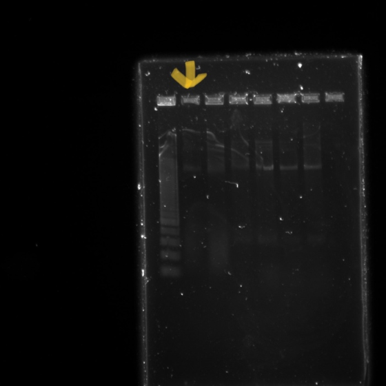
need the answers for questions 2 and 3. My DNA ladder is in lane 2...
I just need the answers to questions 2 and 3. My DNA ladder is in lane...
 I just need the answers to questions 2 and 3. My DNA ladder is
in lane 2 with the yellow arrow pointing to it. Thanks!
Part 2: Gel purification and ration Gel Slice and PCR Product Preparation modified from IBSci.com instructions for gel and PCR clean-up system A. Dissolving the Gel Slice 1. Following electrophoresis, excise DNA band from gel and place gel slice in a 1.5ml microcentrifuge tube. 1b. Use an analytical balance to weigh gel slice. Record weight...
I just need the answers to questions 2 and 3. My DNA ladder is
in lane 2 with the yellow arrow pointing to it. Thanks!
Part 2: Gel purification and ration Gel Slice and PCR Product Preparation modified from IBSci.com instructions for gel and PCR clean-up system A. Dissolving the Gel Slice 1. Following electrophoresis, excise DNA band from gel and place gel slice in a 1.5ml microcentrifuge tube. 1b. Use an analytical balance to weigh gel slice. Record weight...
Question 1. 1 2 3 4 5 6 7 8 Lane Name/Code Ladder 1B | 2B...
 Question 1. 1 2 3 4 5 6 7 8 Lane Name/Code Ladder 1B | 2B 2 DNA Sample/Treatment DNA ladder Digest Digest Digest Digest Digest Digest 3B 4B 5B Negative Figure 1: 1% Super buffer agarose gel electrophoresis of restriction digest of the plasmid containing the gdhA gene insert. Based on the information above answer the following questions? 1. What ladder size used? 2. What are the two top bands and bottom bands representing? 3. Explain why the observed...
14. Like a ladder. Circular DNA from the SV40 virus was isolated and subjected to gel...
14. Like a ladder. Circular DNA from the SV40 virus was isolated and subjected to gel electrophoresis. The results are shown in lane A (the control) of the adjoining gel patterns. (a) Why does the DNA separate in agarose gel electrophoresis? How does the DNA in each band differ? (b) What types of DNA do the various bands represent? (c) What is the significance of the fact that more of the DNA is in slower-moving forms?
You isolate plasmid DNA from bacteria (Questions 7-10) 7) A plasmid is an extrachromosomal circul...
 10 please and 7
You isolate plasmid DNA from bacteria (Questions 7-10) 7) A plasmid is an extrachromosomal circular DNA frequently found in prokaryotes. Aside from being smaller, how is it different from the prokaryotic genome? You place equal amounts of plasmid DNA in 4 different tubes and incubate the DNA with increasing amounts of the enzyme topoisomerase I for 1 hour (0 enzyme units 0.25 enzyme units, 0.5 enzyme units and 1 enzyme unit). You then analyze the plasmid...
10 please and 7
You isolate plasmid DNA from bacteria (Questions 7-10) 7) A plasmid is an extrachromosomal circular DNA frequently found in prokaryotes. Aside from being smaller, how is it different from the prokaryotic genome? You place equal amounts of plasmid DNA in 4 different tubes and incubate the DNA with increasing amounts of the enzyme topoisomerase I for 1 hour (0 enzyme units 0.25 enzyme units, 0.5 enzyme units and 1 enzyme unit). You then analyze the plasmid...
Table 8-3. Interpretation of each lane on the gel. For Lanes 1-8, indicate the size of every DNA ...
 HELP PLEASE! at least with some examples.
size of dna ladder- 1.8cm
We were unable to transcribe this imageTable 8-3. Interpretation of each lane on the gel. For Lanes 1-8, indicate the size of every DNA band on the gel by interpolating values from your standard curve. There may be multiple bands per lane.For EACH band, identify size (interpolated from the standard curve you constructed), identity, shape, and topology DISTANCE EACH BAND IN THE LANE H AS MIGRATED SIzE OF...
HELP PLEASE! at least with some examples.
size of dna ladder- 1.8cm
We were unable to transcribe this imageTable 8-3. Interpretation of each lane on the gel. For Lanes 1-8, indicate the size of every DNA band on the gel by interpolating values from your standard curve. There may be multiple bands per lane.For EACH band, identify size (interpolated from the standard curve you constructed), identity, shape, and topology DISTANCE EACH BAND IN THE LANE H AS MIGRATED SIzE OF...
While your gel is running, discuss your expected results for each lane on the gel •...
 While your gel is running, discuss your expected results for each lane on the gel • Remove the gel from the apparatus and image on a UV transilluminator. (Remember to only illuminate the UV light when the shield is in place). 3) Using the DNA ladder, estimate the experimental size of all bands observed in the sample lanes. (3pt) Gel Lane Plasmid Number of bands observed Estimated size (bp) of each band 2 (tube 1) A 3 (tube 2) B...
En (2 points) You isolated your mitochondrial DNA in Part I. In step 6, you discard...
 En (2 points) You isolated your mitochondrial DNA in Part I. In step 6, you discard the supernatant, but keep the pellet. In step 15, you discard the pellet, but keep the supernatant. Explain why the pattern is different between the two steps and the consequence of mixing up these two steps. Procedure Part 1: mt DNA Isolation from your cheek cells. Lysis solution is used to breakdown the cells in this step, you will isolate MEONA from cheek cells....
3. The given figure represents the agarose gel electrophoresis results from a restriction digest experiment. Lane...
 3. The given figure represents the agarose gel electrophoresis results from a restriction digest experiment. Lane 1 is a DNA ladder (values are in kb) and Lane 2 is the DNA sample cut by a restriction enzyme. What is the size of the band C in lane 2? - 1111111 About 1.8 kb Cannot tell based on the information provided 500 bp About 2 kb © Less than 1.5 bp -
Actual gels don't have labels. Here, the labels have been removed, but the ladder remains the...
 Actual gels don't have labels. Here, the labels have been removed, but the ladder remains the same as in the previous example. 6. On the gel to the right, write the approximate size of each DNA fragment. Write the sizes next to each appropriate band. 7. Imagine that you have a sample of DNA that contains a single, specific DNA sequence. Before you run your gel, you split your sample into two tubes. You run the DNA from the first...
How did you EXPECT your uncut lane to look in the gel image? What ACTUALLY happened?...
 How did you EXPECT your uncut lane to look in the gel image?
What ACTUALLY happened? What is a plausible explanation if there
was any discrepancy?
How did the observed BclI and EcoRI digest results compare to
the expected results? If they differed, list a potential reason
why?
BclI was incubated at 50°C, while EcoRI was incubated at 37°C.
Why was this necessary? What might you predict to occur if you
accidentally switched the temperatures?
3) Using the DNA ladder,...
How did you EXPECT your uncut lane to look in the gel image?
What ACTUALLY happened? What is a plausible explanation if there
was any discrepancy?
How did the observed BclI and EcoRI digest results compare to
the expected results? If they differed, list a potential reason
why?
BclI was incubated at 50°C, while EcoRI was incubated at 37°C.
Why was this necessary? What might you predict to occur if you
accidentally switched the temperatures?
3) Using the DNA ladder,...
 I just need the answers to questions 2 and 3. My DNA ladder is
in lane 2 with the yellow arrow pointing to it. Thanks!
Part 2: Gel purification and ration Gel Slice and PCR Product Preparation modified from IBSci.com instructions for gel and PCR clean-up system A. Dissolving the Gel Slice 1. Following electrophoresis, excise DNA band from gel and place gel slice in a 1.5ml microcentrifuge tube. 1b. Use an analytical balance to weigh gel slice. Record weight...
I just need the answers to questions 2 and 3. My DNA ladder is
in lane 2 with the yellow arrow pointing to it. Thanks!
Part 2: Gel purification and ration Gel Slice and PCR Product Preparation modified from IBSci.com instructions for gel and PCR clean-up system A. Dissolving the Gel Slice 1. Following electrophoresis, excise DNA band from gel and place gel slice in a 1.5ml microcentrifuge tube. 1b. Use an analytical balance to weigh gel slice. Record weight...
 Question 1. 1 2 3 4 5 6 7 8 Lane Name/Code Ladder 1B | 2B 2 DNA Sample/Treatment DNA ladder Digest Digest Digest Digest Digest Digest 3B 4B 5B Negative Figure 1: 1% Super buffer agarose gel electrophoresis of restriction digest of the plasmid containing the gdhA gene insert. Based on the information above answer the following questions? 1. What ladder size used? 2. What are the two top bands and bottom bands representing? 3. Explain why the observed...
 10 please and 7
You isolate plasmid DNA from bacteria (Questions 7-10) 7) A plasmid is an extrachromosomal circular DNA frequently found in prokaryotes. Aside from being smaller, how is it different from the prokaryotic genome? You place equal amounts of plasmid DNA in 4 different tubes and incubate the DNA with increasing amounts of the enzyme topoisomerase I for 1 hour (0 enzyme units 0.25 enzyme units, 0.5 enzyme units and 1 enzyme unit). You then analyze the plasmid...
10 please and 7
You isolate plasmid DNA from bacteria (Questions 7-10) 7) A plasmid is an extrachromosomal circular DNA frequently found in prokaryotes. Aside from being smaller, how is it different from the prokaryotic genome? You place equal amounts of plasmid DNA in 4 different tubes and incubate the DNA with increasing amounts of the enzyme topoisomerase I for 1 hour (0 enzyme units 0.25 enzyme units, 0.5 enzyme units and 1 enzyme unit). You then analyze the plasmid...
 HELP PLEASE! at least with some examples.
size of dna ladder- 1.8cm
We were unable to transcribe this imageTable 8-3. Interpretation of each lane on the gel. For Lanes 1-8, indicate the size of every DNA band on the gel by interpolating values from your standard curve. There may be multiple bands per lane.For EACH band, identify size (interpolated from the standard curve you constructed), identity, shape, and topology DISTANCE EACH BAND IN THE LANE H AS MIGRATED SIzE OF...
HELP PLEASE! at least with some examples.
size of dna ladder- 1.8cm
We were unable to transcribe this imageTable 8-3. Interpretation of each lane on the gel. For Lanes 1-8, indicate the size of every DNA band on the gel by interpolating values from your standard curve. There may be multiple bands per lane.For EACH band, identify size (interpolated from the standard curve you constructed), identity, shape, and topology DISTANCE EACH BAND IN THE LANE H AS MIGRATED SIzE OF...
 While your gel is running, discuss your expected results for each lane on the gel • Remove the gel from the apparatus and image on a UV transilluminator. (Remember to only illuminate the UV light when the shield is in place). 3) Using the DNA ladder, estimate the experimental size of all bands observed in the sample lanes. (3pt) Gel Lane Plasmid Number of bands observed Estimated size (bp) of each band 2 (tube 1) A 3 (tube 2) B...
 En (2 points) You isolated your mitochondrial DNA in Part I. In step 6, you discard the supernatant, but keep the pellet. In step 15, you discard the pellet, but keep the supernatant. Explain why the pattern is different between the two steps and the consequence of mixing up these two steps. Procedure Part 1: mt DNA Isolation from your cheek cells. Lysis solution is used to breakdown the cells in this step, you will isolate MEONA from cheek cells....
 3. The given figure represents the agarose gel electrophoresis results from a restriction digest experiment. Lane 1 is a DNA ladder (values are in kb) and Lane 2 is the DNA sample cut by a restriction enzyme. What is the size of the band C in lane 2? - 1111111 About 1.8 kb Cannot tell based on the information provided 500 bp About 2 kb © Less than 1.5 bp -
 Actual gels don't have labels. Here, the labels have been removed, but the ladder remains the same as in the previous example. 6. On the gel to the right, write the approximate size of each DNA fragment. Write the sizes next to each appropriate band. 7. Imagine that you have a sample of DNA that contains a single, specific DNA sequence. Before you run your gel, you split your sample into two tubes. You run the DNA from the first...
 How did you EXPECT your uncut lane to look in the gel image?
What ACTUALLY happened? What is a plausible explanation if there
was any discrepancy?
How did the observed BclI and EcoRI digest results compare to
the expected results? If they differed, list a potential reason
why?
BclI was incubated at 50°C, while EcoRI was incubated at 37°C.
Why was this necessary? What might you predict to occur if you
accidentally switched the temperatures?
3) Using the DNA ladder,...
How did you EXPECT your uncut lane to look in the gel image?
What ACTUALLY happened? What is a plausible explanation if there
was any discrepancy?
How did the observed BclI and EcoRI digest results compare to
the expected results? If they differed, list a potential reason
why?
BclI was incubated at 50°C, while EcoRI was incubated at 37°C.
Why was this necessary? What might you predict to occur if you
accidentally switched the temperatures?
3) Using the DNA ladder,...
Most questions answered within 3 hours.
-
Cisco packet tracer
Q1) Do you get any changes of IP address when packet is
traversing...
asked 28 minutes ago -
What is the pressure inside a 33.0 L container holding 106.4 kg
of argon gas at...
asked 1 hour ago -
Question no 2
A housekeeping support department budgets its costs at
SR 40,000 per month plus...
asked 1 hour ago -
A 1400Kg sports car accelerates from rest to 90km/h in 7.0s.
What is the average power...
asked 1 hour ago -
For the following reaction, 0.128 moles of
potassium hydrogen sulfateare mixed with
0.504 moles of potassium...
asked 5 hours ago -
1. What is the present value of $400, three years in the future
if the interest...
asked 6 hours ago -
The labor force minus the number of employed equals the number
of unemployed.
a. True
b....
asked 8 hours ago -
Determine the mass in units of grams [g] of 0.49 moles [mol]
of a new fictitious...
asked 8 hours ago -
A horizontal mass of M=5kg is on a spring and stretched to
x=0.5m when released from...
asked 9 hours ago -
26 of 50
"I have worked at the Arizona Humane Society for ten years, and
have...
asked 10 hours ago -
Compare and contrast zero based budgeting and incremental (or
base year) budgeting.
asked 10 hours ago -
4 pts 10. Which of the following hypothesis would be MOST
difficult to test experimentally? Group...
asked 10 hours ago