
Homework Answers
Add Answer to:
QUESTION 8 Final images appear in 3-D when specimens are viewed with these microscopes:- O A....
QUESTION 9 Which of the following microscopes produce flat images by utilizing light in the non-visual...
 QUESTION 9 Which of the following microscopes produce flat images by utilizing light in the non-visual spectrum to excite molecules within the specimens to produce visual light? Darkfield Microscope Confocal Microscope Phase Contrast Microscope Fluorescent Microscope Brightfield Microscope Electron Microscope QUESTION 10 Which type of microscope does not use glass lenses or mirrors in order to focus and view a specimen? Brightfield Microscope Darkfield Microscope Confocal Microscope Phase Contrast Microscope Electron Microscope Fluorescent Microscope
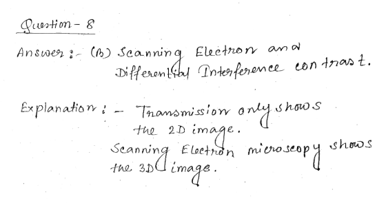
What type of microscope (see below) would you use to greatly magnify a tiny 3-D structure?...
What type of microscope (see below) would you use to greatly magnify a tiny 3-D structure? Why? Bright field, dark field, phase contrast, differential-interference, fluorescence, conofocal, transmission electron, or scanning electron
QUESTION 3 Which of the following light microscopes require staining to view details of a fixed...
 QUESTION 3 Which of the following light microscopes require staining to view details of a fixed specimen on a light background? Inverted Microscope All of these Phase Contrast Microscope Confocal Microscope Darkfield Microscope Brightfield Microscope QUESTION 4 Which of the following would be the proper use of a microscope? Select all that apply. Move the microscope by carrying it by the arm and the base. After use turn off the light and unplug the microscope After using the oil immersion...
Question 1 Which electron microscopes type offers higher resolution? Atomic force microscopy. b. Scanning electron microscopy....
 Question 1 Which electron microscopes type offers higher resolution? Atomic force microscopy. b. Scanning electron microscopy. Phase contrast microscopy. Transmission electron microscopy. A Moving to the next question prevents changes to this answer. Type here to search Which of these statements is correct for gram staining? Which of these statements is correct for gram staining? Carbolfuchsin-mordant-alcohol-malachite green. Ob Methylene blue-mordant-alcohol-crystal violet. Carbolfuchsin-alcohol-methylene blue. d Crystal violet-alcohol-mordant-methylene blue. Crystal violet-mordant-alcohol-safranin.
121. A graduate student is studying a pathological bacterial strain. As a part of a project...
 121. A graduate student is studying a pathological bacterial strain. As a part of a project to develop an antibiotic against the bacterial strain, the student needs to determine the number of proteins present in the ribosome. The best technique for investigating this question is: A. Cell homogenization and centrifugation B. Phase contrast microscopy C. Brightfield microscopy D. Scanning electron microscopy E. Transmission electron microscopy
2)Which of the A)peptidoglycan B)cellulose C)glycogen D)chitin 3 Which of the following is true of fluorescence...
 2)Which of the A)peptidoglycan B)cellulose C)glycogen D)chitin 3 Which of the following is true of fluorescence microscopy? A) Fluorescence microscopy is best at viewing rounded, thicker specimens B) Fluorescent light is emitted throughout the specimen due to fluorescent dyes conjugated to antibodies. C) Fluorescence microscopy is used to view dead specimens only. D) Fluorescence microscopy presents images in three dimensions. E) Fluorescence microscopy is able to overcome problems encountered with using confocal scanning microscopy. 4)While synthesizing a new green pigment,...
Immunocytochemistry is the method of visualization of proteins in cells with the use of antibody that...
 Immunocytochemistry is the method of visualization of proteins in cells with the use of antibody that recognize specific molecules. Recently the visualization of these antibody-bound molecules is realized with the imaging of fluorescently-labeled antibodies against these receptors. Which type of microscopy would be used for this study? 0. Confocal microscopy O. Phase-contrast microscopy O. Differential interference contrast microscopy Electron phase-contrast microscopy You are working on a project that involves an imaging of rough endoplasmic reticulum shown here. What microscope was...
1. Fill out the following table by indicating which general technique (light microscopy (LM) or electron...
 1. Fill out the following table by indicating which general technique (light microscopy (LM) or electron microscopy (EM]) could be used to observe each structure or phenomenon. Put "no" in the box if the technique could not be used. If light microscopy can be used, name one technique (bright-field, phase-contrast, fluorescence, etc.) that you think would be effective. You will find some useful information in Appendix 1 of this manual and Chapter 18 of your textbook. Structure or phenomenon Could...
QUESTION 32 Nearsightedness can result when the eyeball is elongated sach that the images of a...
 QUESTION 32 Nearsightedness can result when the eyeball is elongated sach that the images of a nearby objects are focused behind the retina b. distant objects are focused in front of the retina. O c. nearby objects are magnified to twice their original size. d. distant objects are shrunk to less than their original size.
QUESTION 1 Specimens for cold agglutinins must be: A. transported on ice © B. kept warm...
 QUESTION 1 Specimens for cold agglutinins must be: A. transported on ice © B. kept warm O C. drawn in a green top tube D. processed in a refrigerated centrifuge QUESTION 2 The fasting specimen for a GTT is drawn at 0700, and the patient finishes drinking the glucola A. 0800 OB. 0815 C. 0845 D. 0915 QUESTION 3 When collecting units of donor blood, the most frequently used needle gauge is: A. 26 B. 23
 QUESTION 9 Which of the following microscopes produce flat images by utilizing light in the non-visual spectrum to excite molecules within the specimens to produce visual light? Darkfield Microscope Confocal Microscope Phase Contrast Microscope Fluorescent Microscope Brightfield Microscope Electron Microscope QUESTION 10 Which type of microscope does not use glass lenses or mirrors in order to focus and view a specimen? Brightfield Microscope Darkfield Microscope Confocal Microscope Phase Contrast Microscope Electron Microscope Fluorescent Microscope
 QUESTION 3 Which of the following light microscopes require staining to view details of a fixed specimen on a light background? Inverted Microscope All of these Phase Contrast Microscope Confocal Microscope Darkfield Microscope Brightfield Microscope QUESTION 4 Which of the following would be the proper use of a microscope? Select all that apply. Move the microscope by carrying it by the arm and the base. After use turn off the light and unplug the microscope After using the oil immersion...
 Question 1 Which electron microscopes type offers higher resolution? Atomic force microscopy. b. Scanning electron microscopy. Phase contrast microscopy. Transmission electron microscopy. A Moving to the next question prevents changes to this answer. Type here to search Which of these statements is correct for gram staining? Which of these statements is correct for gram staining? Carbolfuchsin-mordant-alcohol-malachite green. Ob Methylene blue-mordant-alcohol-crystal violet. Carbolfuchsin-alcohol-methylene blue. d Crystal violet-alcohol-mordant-methylene blue. Crystal violet-mordant-alcohol-safranin.
 121. A graduate student is studying a pathological bacterial strain. As a part of a project to develop an antibiotic against the bacterial strain, the student needs to determine the number of proteins present in the ribosome. The best technique for investigating this question is: A. Cell homogenization and centrifugation B. Phase contrast microscopy C. Brightfield microscopy D. Scanning electron microscopy E. Transmission electron microscopy
 2)Which of the A)peptidoglycan B)cellulose C)glycogen D)chitin 3 Which of the following is true of fluorescence microscopy? A) Fluorescence microscopy is best at viewing rounded, thicker specimens B) Fluorescent light is emitted throughout the specimen due to fluorescent dyes conjugated to antibodies. C) Fluorescence microscopy is used to view dead specimens only. D) Fluorescence microscopy presents images in three dimensions. E) Fluorescence microscopy is able to overcome problems encountered with using confocal scanning microscopy. 4)While synthesizing a new green pigment,...
 Immunocytochemistry is the method of visualization of proteins in cells with the use of antibody that recognize specific molecules. Recently the visualization of these antibody-bound molecules is realized with the imaging of fluorescently-labeled antibodies against these receptors. Which type of microscopy would be used for this study? 0. Confocal microscopy O. Phase-contrast microscopy O. Differential interference contrast microscopy Electron phase-contrast microscopy You are working on a project that involves an imaging of rough endoplasmic reticulum shown here. What microscope was...
 1. Fill out the following table by indicating which general technique (light microscopy (LM) or electron microscopy (EM]) could be used to observe each structure or phenomenon. Put "no" in the box if the technique could not be used. If light microscopy can be used, name one technique (bright-field, phase-contrast, fluorescence, etc.) that you think would be effective. You will find some useful information in Appendix 1 of this manual and Chapter 18 of your textbook. Structure or phenomenon Could...
 QUESTION 32 Nearsightedness can result when the eyeball is elongated sach that the images of a nearby objects are focused behind the retina b. distant objects are focused in front of the retina. O c. nearby objects are magnified to twice their original size. d. distant objects are shrunk to less than their original size.
 QUESTION 1 Specimens for cold agglutinins must be: A. transported on ice © B. kept warm O C. drawn in a green top tube D. processed in a refrigerated centrifuge QUESTION 2 The fasting specimen for a GTT is drawn at 0700, and the patient finishes drinking the glucola A. 0800 OB. 0815 C. 0845 D. 0915 QUESTION 3 When collecting units of donor blood, the most frequently used needle gauge is: A. 26 B. 23
Most questions answered within 3 hours.
-
Living in a group could bring several disadvantages to an
individual. What are some of the...
asked 2 minutes ago -
Complete and balance the following reactions. In case of no
reaction occurring write NR.
Mix #1:...
asked 8 minutes ago -
If an economy consumes 75% of any increase in income, then an
increase in autonomous investment...
asked 11 minutes ago -
A shotputter throws the shot with an initial speed of 15.8 m/s
at a 38.0 ∘...
asked 27 minutes ago -
Debra and Merina sell electronic equipment and supplies through
their partnership. They wish to expand their...
asked 28 minutes ago -
How does a linear regression allow you to better estimate
trends, costs, and other factors in...
asked 36 minutes ago -
1. (15%) Describe the difference between a pull (Kanban), push
and CONWIP production systems.
asked 32 minutes ago -
QUESTION 5
The total area under the Z distribution curve is equal to:
a.
10
b....
asked 41 minutes ago -
Using Python
The variables x and y refer to numbers. Write a code segment
that prompts...
asked 55 minutes ago -
If
the coefficient of static friction between a box and the floor is
0.35 with what...
asked 57 minutes ago -
A die is designed to punch holes with a radius of 1.08 10-2 m in
a...
asked 1 hour ago -
government can increase import through
a. export subsidies
b. tax breaks
c. increase import tax
d....
asked 1 hour ago