You are using a ChIP assay to study the positioning of a DNA-binding transcription factor
(Dbp1p) on transcribed genes. You fragment DNA from wild type cells and immunoprecipitate
Dbp1p with an antibody to it. Then you PCR amplify the DNA associated with Dbp1p using
different sets of primers for a particular gene (G3PD). One set of primers was specific for the
TATA box region of this gene, (lane 1) another pair of primers amplified the 3’ end of the open
reading frame for G3PD(lane 2); and a 3rd set of primers amplified the region just beyond the
polyadenylation site downstream of the open reading frame (lane 3). PCR products from
chromatin immunoprecipitation (right gel below) were compared to PCR reactions using input
chromatin without immunoprecipitating (left gel below). You also included a PCR reaction with
primers specific for a nontranscribed region (lowest band on all the gels below). Results are
shown.
A) Using a standard line drawing, show the G3PD gene (TATA, Trxn start, AUG, Stop
codon, polyA site) and draw the 4 primer pairs from above at the respective positions that
they amplify (to scale approximately).
B) For the input PCR reaction (left), explain what each of the bands represents. Why are the
bands different sizes (different mobilities) in each lane?
C) Explain why all the bands are of the same intensity for the input PCR reactions
D) What is the conclusion from the ChIP? Explain. What lanes/controls support your ideas?

Homework Answers
1.
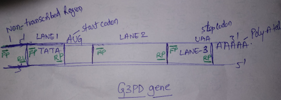
2.The band in LANE 1 represents TATA ( promoter region) may be having the low moleculat weight having much mobility than the remaining two, LANE 2 represents 3' end of open reading frame having more molecular weight than the remaining two and LANE 3 represnts next to 3' end of open reading frame i.e before polyadenylation tail having the medium molecular weight than the remaining two electrophoresed inbetween the two bands. Difference in molecular weights caused them to mobilize in three different patterns. The bands in the lower panel are from untranscribed region having low molecular weight than the three and was a positive control.
3. All bands in the input reaction are having same intensity because all of them are having high expression levels more of less equal so they are having same intensity.
4. The transcription factor of 3' end of open reading frame is expressed in high levels compared with the remaining. Equal expression in the lower panel ( in the left and right pcr reactions) indicate equal amounts of loaded DNA (like house keeping gene to determine the equal amounts of loaded DNA).
Add Answer to:
You are using a ChIP assay to study the positioning of a
DNA-binding transcription factor
(Dbp1p)...
You PCR amplify a 500 bp (base pairs) piece of DNA that has diagnostic value in...
 You PCR amplify a 500 bp (base pairs) piece of DNA that has
diagnostic value in determining whether a patient has a mutation
within a specific DNA region. You know that this DNA segment of the
“normal” gene does not include an EcoRI restriction site; but the
mutated DNA segment of the same gene contains an EcoRI restriction
site due to a point mutation at the 100th bp from the 5’ DNA end.
After PCR amplification, you subject your DNA...
You PCR amplify a 500 bp (base pairs) piece of DNA that has
diagnostic value in determining whether a patient has a mutation
within a specific DNA region. You know that this DNA segment of the
“normal” gene does not include an EcoRI restriction site; but the
mutated DNA segment of the same gene contains an EcoRI restriction
site due to a point mutation at the 100th bp from the 5’ DNA end.
After PCR amplification, you subject your DNA...
EXPLAIN each answer thoroughly. There are two common goals when using PCR to amplify DNA. A....
EXPLAIN each answer thoroughly. There are two common goals when using PCR to amplify DNA. A. Make lots of copies of a specific DNA sequence to use in cloning (preparative PCR) B. Detect the presence or relative amount of a specific gene under varying conditions (analytical PCR) *Remember: PCR is very similiar to DNA replication, but uses a DNA polymerase to amplify only specific parts of DNA sequence based on the sequence of the primers. 3. For which goal (A...
You are interested in the interactions of histone deacetylase (HDAC) with nucleosomes. Using a DNA template...
 You are interested in the interactions of histone deacetylase
(HDAC) with nucleosomes. Using a DNA template that is end labelled
with 32P and has two nucleosomes already bound to it, you perform a
gel-shift experiment with HDAC and the chromatin from above. You
also assemble chromatin with nucleosomes that have been methylated
at a particular amino acid residue of Histone H4. The results are
below. Based on the data, (A) explain the autoradiogram below –
what are all the bands?...
You are interested in the interactions of histone deacetylase
(HDAC) with nucleosomes. Using a DNA template that is end labelled
with 32P and has two nucleosomes already bound to it, you perform a
gel-shift experiment with HDAC and the chromatin from above. You
also assemble chromatin with nucleosomes that have been methylated
at a particular amino acid residue of Histone H4. The results are
below. Based on the data, (A) explain the autoradiogram below –
what are all the bands?...
2. PCR amplification of the TAS2R38 gene a. The number of copies of the 303 bp sequence grows exp...
 2. PCR amplification of the TAS2R38 gene a. The number of copies of the 303 bp sequence grows exponentially (1-2-4-8-etc) after each cycle. The number of cycles we used is on page 97. What is the number of copies of the 303 bp fragment that will theoretically be present at the end of our reaction? b. Denaturation of the 303 bp segment of the TAS2R38 gene is a critical first step in the PCR perties of a DNA segment that...
QUESTION 1: You are inserting a gene into an MCS found within the LacZ gene. Using...
QUESTION 1: You are inserting a gene into an MCS found within the LacZ gene. Using blue/white colony selection, why could you assume that white colonies have modified plasmids? a. A blue colony means the LacZ reading-frame was disrupted b. A blue colony means your gene has mutations c. A white colony means the LacZ reading-frame is intact d. A white colony means the LacZ reading-frame was disrupted QUESTION 2: You are performing a PCR using primers with a sequence perfectly...
can someone explain how to answer this with reasons why 30-45 mins after estrogen addition: Acchyiase...
 can someone explain how to answer this with reasons why
30-45 mins after estrogen addition: Acchyiase (HAT, odde auhye 60-90 mins after estrogen addition: Reauitment ef Poy to open 7. T identify the Gene X DNA element responsible for regulation by a Growth Fagor, you perform a linker scanning experiment in which you mutate 10 bp Tegions centered around positions -205 to-5 in the Gene X promoter/promoter- proximal region (wt wild-type promoter). The promoter variants were tested for their ability...
can someone explain how to answer this with reasons why
30-45 mins after estrogen addition: Acchyiase (HAT, odde auhye 60-90 mins after estrogen addition: Reauitment ef Poy to open 7. T identify the Gene X DNA element responsible for regulation by a Growth Fagor, you perform a linker scanning experiment in which you mutate 10 bp Tegions centered around positions -205 to-5 in the Gene X promoter/promoter- proximal region (wt wild-type promoter). The promoter variants were tested for their ability...
In DNA sequencing, ddNTPs differ from dNTPs in that ddNTPs are lacking a OH at the...
 In DNA sequencing, ddNTPs differ from dNTPs in that ddNTPs are
lacking a OH at the
a.
2' carbon
b.
5' carbon
c.
3' carbon
please answer as many as you can!! I am low on questions and
could use the help!
Mother || Child II Male 1 Male 2 Results from a paternity test using DNA fingerprinting is shown. DNA was isolated from a mother, her child and 2 potential fathers. Primers designed to amplify different satellite DNA regions...
In DNA sequencing, ddNTPs differ from dNTPs in that ddNTPs are
lacking a OH at the
a.
2' carbon
b.
5' carbon
c.
3' carbon
please answer as many as you can!! I am low on questions and
could use the help!
Mother || Child II Male 1 Male 2 Results from a paternity test using DNA fingerprinting is shown. DNA was isolated from a mother, her child and 2 potential fathers. Primers designed to amplify different satellite DNA regions...
The PCR was a success and your target region of 770 bp in length has been...
 The PCR was a success and your target region of 770 bp in length has been amplified. You now plan to digest the DNA amplicon with the restriction enzyme Eael, and clone the resulting longest fragment it into the Eael site of the 5 kb plasmid diagrammed below. 770 bp BamHI 1 200 EcoRI 800 EcoRI 4000 1000 5 kb O /1000 2000 2000 Faal You purify your recombinant plasmid from bacterial cells, and run the plasmid (uncut. or not...
You are using PCR to amplify a 300 bp target sequence, a portion of Gene X,...
You are using PCR to amplify a 300 bp target sequence, a portion of Gene X, from human genomic DNA isolated from patients' blood samples. The instructions for this procedure tell you to include Samples A and B, whose contents are listed below, with each batch of patient samples that you run. Ingredients Sample A Sample B 10x PCR Buffer (Tris,KCI,MgCl2,BSA) 5 mL 5 mL H2O 37.8mL 38.8mL dNTP's 3 mL 3 mL Taq DNA polymerase 0.2 mL 0.2 mL...
En (2 points) You isolated your mitochondrial DNA in Part I. In step 6, you discard...
 En (2 points) You isolated your mitochondrial DNA in Part I. In step 6, you discard the supernatant, but keep the pellet. In step 15, you discard the pellet, but keep the supernatant. Explain why the pattern is different between the two steps and the consequence of mixing up these two steps. Procedure Part 1: mt DNA Isolation from your cheek cells. Lysis solution is used to breakdown the cells in this step, you will isolate MEONA from cheek cells....
 You PCR amplify a 500 bp (base pairs) piece of DNA that has
diagnostic value in determining whether a patient has a mutation
within a specific DNA region. You know that this DNA segment of the
“normal” gene does not include an EcoRI restriction site; but the
mutated DNA segment of the same gene contains an EcoRI restriction
site due to a point mutation at the 100th bp from the 5’ DNA end.
After PCR amplification, you subject your DNA...
You PCR amplify a 500 bp (base pairs) piece of DNA that has
diagnostic value in determining whether a patient has a mutation
within a specific DNA region. You know that this DNA segment of the
“normal” gene does not include an EcoRI restriction site; but the
mutated DNA segment of the same gene contains an EcoRI restriction
site due to a point mutation at the 100th bp from the 5’ DNA end.
After PCR amplification, you subject your DNA...
 You are interested in the interactions of histone deacetylase
(HDAC) with nucleosomes. Using a DNA template that is end labelled
with 32P and has two nucleosomes already bound to it, you perform a
gel-shift experiment with HDAC and the chromatin from above. You
also assemble chromatin with nucleosomes that have been methylated
at a particular amino acid residue of Histone H4. The results are
below. Based on the data, (A) explain the autoradiogram below –
what are all the bands?...
You are interested in the interactions of histone deacetylase
(HDAC) with nucleosomes. Using a DNA template that is end labelled
with 32P and has two nucleosomes already bound to it, you perform a
gel-shift experiment with HDAC and the chromatin from above. You
also assemble chromatin with nucleosomes that have been methylated
at a particular amino acid residue of Histone H4. The results are
below. Based on the data, (A) explain the autoradiogram below –
what are all the bands?...
 2. PCR amplification of the TAS2R38 gene a. The number of copies of the 303 bp sequence grows exponentially (1-2-4-8-etc) after each cycle. The number of cycles we used is on page 97. What is the number of copies of the 303 bp fragment that will theoretically be present at the end of our reaction? b. Denaturation of the 303 bp segment of the TAS2R38 gene is a critical first step in the PCR perties of a DNA segment that...
 can someone explain how to answer this with reasons why
30-45 mins after estrogen addition: Acchyiase (HAT, odde auhye 60-90 mins after estrogen addition: Reauitment ef Poy to open 7. T identify the Gene X DNA element responsible for regulation by a Growth Fagor, you perform a linker scanning experiment in which you mutate 10 bp Tegions centered around positions -205 to-5 in the Gene X promoter/promoter- proximal region (wt wild-type promoter). The promoter variants were tested for their ability...
can someone explain how to answer this with reasons why
30-45 mins after estrogen addition: Acchyiase (HAT, odde auhye 60-90 mins after estrogen addition: Reauitment ef Poy to open 7. T identify the Gene X DNA element responsible for regulation by a Growth Fagor, you perform a linker scanning experiment in which you mutate 10 bp Tegions centered around positions -205 to-5 in the Gene X promoter/promoter- proximal region (wt wild-type promoter). The promoter variants were tested for their ability...
 In DNA sequencing, ddNTPs differ from dNTPs in that ddNTPs are
lacking a OH at the
a.
2' carbon
b.
5' carbon
c.
3' carbon
please answer as many as you can!! I am low on questions and
could use the help!
Mother || Child II Male 1 Male 2 Results from a paternity test using DNA fingerprinting is shown. DNA was isolated from a mother, her child and 2 potential fathers. Primers designed to amplify different satellite DNA regions...
In DNA sequencing, ddNTPs differ from dNTPs in that ddNTPs are
lacking a OH at the
a.
2' carbon
b.
5' carbon
c.
3' carbon
please answer as many as you can!! I am low on questions and
could use the help!
Mother || Child II Male 1 Male 2 Results from a paternity test using DNA fingerprinting is shown. DNA was isolated from a mother, her child and 2 potential fathers. Primers designed to amplify different satellite DNA regions...
 The PCR was a success and your target region of 770 bp in length has been amplified. You now plan to digest the DNA amplicon with the restriction enzyme Eael, and clone the resulting longest fragment it into the Eael site of the 5 kb plasmid diagrammed below. 770 bp BamHI 1 200 EcoRI 800 EcoRI 4000 1000 5 kb O /1000 2000 2000 Faal You purify your recombinant plasmid from bacterial cells, and run the plasmid (uncut. or not...
 En (2 points) You isolated your mitochondrial DNA in Part I. In step 6, you discard the supernatant, but keep the pellet. In step 15, you discard the pellet, but keep the supernatant. Explain why the pattern is different between the two steps and the consequence of mixing up these two steps. Procedure Part 1: mt DNA Isolation from your cheek cells. Lysis solution is used to breakdown the cells in this step, you will isolate MEONA from cheek cells....
Most questions answered within 3 hours.
-
Identify which one of these systems can be considered as
constituting an isolated system.
Two marbles...
asked 8 seconds from now -
10. Explain what is meant by ‘mutually exclusive projects’ and
why it is generally a bad...
asked 10 minutes ago -
Create a powershell script to create a user named: Week10User
(provide whatever other details that may...
asked 4 minutes ago -
QUESTION 10
Which of the following is not true of nonprofit Community Health
Centers that qualify...
asked 5 minutes ago -
Suppose that you wanted to purchase a product and sell it at
your online store. You...
asked 10 minutes ago -
The coefficient of determination R2 in a simple
regression model,
Group of answer choices
a) measures...
asked 11 minutes ago -
Explain why some heterotrophs can be described as both a
primary consumer and a secondary consumer....
asked 15 minutes ago -
Explain the constitutional foundations for federalism.
asked 27 minutes ago -
Perform an internet search and answer the following questions:
Why should a business be concerned with...
asked 36 minutes ago -
Which board(s) has (have) worked to implement fair value
measurement for financial instruments? A. FASB, but...
asked 40 minutes ago -
Consider an element that reaches its first excited state by
absorption of 457.8 nm light.
Determine...
asked 51 minutes ago -
Sandhill Company sells product 2005WSC for $135 per unit. The
cost of one unit of 2005WSC...
asked 46 minutes ago