
Homework Answers
The orbit is the compartment or cavity in which eye is situated. It is basically comprised of seven bones.
The seven bones are as following:
1.) Frontal bone
2.) Sphenoid bone: a.) Greater wing of Sphenoid and b.) Lesser wing
of Sphenoid
3.) Maxillary bone
4.) Zygomatic bone
5.) Ethmoid bone
6.) Palatine bone
7.) Lacrimal bone
Let’s define each term.
1.) Frontal bone: This bone is the part of the
human skull which is found in the forehead region.
2.) Sphenoid bone: It is the bone that helps in
the formation of skull and provides rigidity to it. Greater wing
and lesser wing articulate into formation of sphenoid bone. Another
key feature of this bone is that it joins with almost every bone of
skull.
3.) Maxillary bone: These are the bones which help
in the formation of upper jaw and have their contribution in orbit
formation as well.
4.) Zygomatic bone: Also called as cheek bone has
two extensions formed from maxilla and temporal bone. Both these
extensions contribute in the formation of zygomatic bone.
5.) Ethmoid bone: It is the bone located between
the eyes and helps in the formation of nasal cavity, orbit and
various other parts. It is also very light and spongy bone.
6.) Palatine bone: It has a smaller contribution
to the formation of orbit as well as nasal cavity. It also forms
the hard palate hence the name Palatine.
7.) Lacrimal bone: It is a facial bone that helps
in the formation of orbit. It is very small in size. You might have
heard of lacrimal glands which helps in tear formation. This bone
provides a canal to drain out that tear.
All these bones with their contributions helps to form the orbit.
Add Answer to:
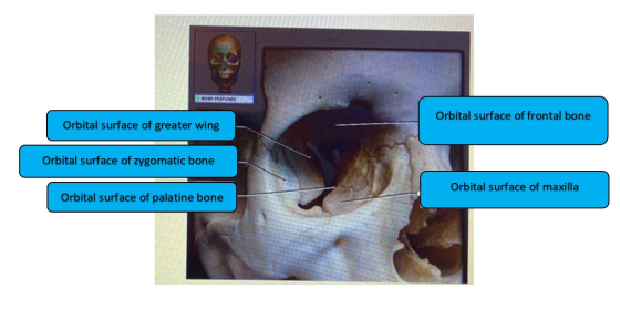
avity Sred Label the bony features of the orbit Orbital surface of greater wing Orbital surface...
Saved omework Assignment Label the bones of the skull in inferior view. Maxilla Palatine bone Zygomatic...
 Saved omework Assignment Label the bones of the skull in inferior view. Maxilla Palatine bone Zygomatic arch Occipital bone Sphenoid bone Greater wing of sphenoid Vomer Temporal bone Parietal bone Reset Zoom
fill in the blank Label the specific bony features of the superior skull. Skull Occipital bone...
 fill in the blank
Label the specific bony features of the superior skull. Skull Occipital bone Sutural bone DONE FEATURES Sagittal suture Parietal bone Frontal bone Coronal suture Lambdoid suture Superior view Reset Zoom
fill in the blank
Label the specific bony features of the superior skull. Skull Occipital bone Sutural bone DONE FEATURES Sagittal suture Parietal bone Frontal bone Coronal suture Lambdoid suture Superior view Reset Zoom
136 Review Sheet 9 2. Using choices from the numbered key to the right, identify all...
 136 Review Sheet 9 2. Using choices from the numbered key to the right, identify all bones and bone markings provided with various leader lines in the two following photographs. A colored dot at the end of a leader line indicates a bone leader lines without a colored dot indicate bone markings. Note that vomer, sphenoid bone, and zygomatic bone will each be labeled twice. Key: 1. alveolar processes 2. carotid canal 3. ethmoid bone (perpendicular plate) 4. external occipital...
vity Savod Label the specific bony features of the skull in lateral view Pronion Lacrimal fossa...
 vity Savod Label the specific bony features of the skull in lateral view Pronion Lacrimal fossa External acoustic meatus Squamous suture Coronal suture Lambdoid suture < Prev 11 of 69 !!! Next > earch hp
ty Label the specific bony features of the skull in inferior view. Formen ovale Foramen magnum...
 ty Label the specific bony features of the skull in inferior view. Formen ovale Foramen magnum Foramen spinosum Lateral plerygoid plate Choana External occipital protuberance Jugular foramen Medial pterygoid plate < Prev 17 of 69 Next > search
dieu. Skeleldi 1 Save Help Save & Label the specific bony features in the superior view...
 dieu. Skeleldi 1 Save Help Save & Label the specific bony features in the superior view of the cranial cavity. Jugular foramen Lesser wing of sphenoid Internal acoustic meatus Hypoglossal canal ences Optic canal Sella turcica Superior orbital fissure Hypophyseal fossa Reset Zoom
Saved wity Label the structures of the bone using the hints provided. Sphenoid bone Pterygoid canal...
 Saved wity Label the structures of the bone using the hints provided. Sphenoid bone Pterygoid canal Optic canal Greater wing Posterior view Lesser wing < Prev 26 of 69 !!! Next > e to search
PRE-LABORATORY WORKSHEET Chapter 8: The Skeletal System: Axial Skeleton These Pre-Laboratory Worksheet questions may be assigned...
 PRE-LABORATORY WORKSHEET Chapter 8: The Skeletal System: Axial Skeleton These Pre-Laboratory Worksheet questions may be assigned by instructors through their connect course. d in com March the description of the bone featurested in column with the appropriate name Column CA I smooth, grooved, pulele process to facet 9 2 small flat shallow, articulating surface B a large amooth and round projection C e troches prominent, rounded epiphysis of a bone 2. Which of the following are bony features that function...
 Saved omework Assignment Label the bones of the skull in inferior view. Maxilla Palatine bone Zygomatic arch Occipital bone Sphenoid bone Greater wing of sphenoid Vomer Temporal bone Parietal bone Reset Zoom
 fill in the blank
Label the specific bony features of the superior skull. Skull Occipital bone Sutural bone DONE FEATURES Sagittal suture Parietal bone Frontal bone Coronal suture Lambdoid suture Superior view Reset Zoom
fill in the blank
Label the specific bony features of the superior skull. Skull Occipital bone Sutural bone DONE FEATURES Sagittal suture Parietal bone Frontal bone Coronal suture Lambdoid suture Superior view Reset Zoom
 136 Review Sheet 9 2. Using choices from the numbered key to the right, identify all bones and bone markings provided with various leader lines in the two following photographs. A colored dot at the end of a leader line indicates a bone leader lines without a colored dot indicate bone markings. Note that vomer, sphenoid bone, and zygomatic bone will each be labeled twice. Key: 1. alveolar processes 2. carotid canal 3. ethmoid bone (perpendicular plate) 4. external occipital...
 vity Savod Label the specific bony features of the skull in lateral view Pronion Lacrimal fossa External acoustic meatus Squamous suture Coronal suture Lambdoid suture < Prev 11 of 69 !!! Next > earch hp
 ty Label the specific bony features of the skull in inferior view. Formen ovale Foramen magnum Foramen spinosum Lateral plerygoid plate Choana External occipital protuberance Jugular foramen Medial pterygoid plate < Prev 17 of 69 Next > search
 dieu. Skeleldi 1 Save Help Save & Label the specific bony features in the superior view of the cranial cavity. Jugular foramen Lesser wing of sphenoid Internal acoustic meatus Hypoglossal canal ences Optic canal Sella turcica Superior orbital fissure Hypophyseal fossa Reset Zoom
 Saved wity Label the structures of the bone using the hints provided. Sphenoid bone Pterygoid canal Optic canal Greater wing Posterior view Lesser wing < Prev 26 of 69 !!! Next > e to search
 PRE-LABORATORY WORKSHEET Chapter 8: The Skeletal System: Axial Skeleton These Pre-Laboratory Worksheet questions may be assigned by instructors through their connect course. d in com March the description of the bone featurested in column with the appropriate name Column CA I smooth, grooved, pulele process to facet 9 2 small flat shallow, articulating surface B a large amooth and round projection C e troches prominent, rounded epiphysis of a bone 2. Which of the following are bony features that function...
Most questions answered within 3 hours.
-
Identify one individual who, in your opinion, is an excellent
leader. List the qualities that this...
asked 1 minute from now -
MAN3240 Organizational Behavior
In one to two paragraphs
6.) How can understanding emotions make me more...
asked 1 minute ago -
For the data set shown below, complete parts (a) through (d)
below. x 3 4 5...
asked 4 minutes ago -
A university administrator working in student housing wants to
determine if the percentage of students residing...
asked 18 minutes ago -
3). Describe human population growth that has occurred in the
past 400 years. Use terms learned...
asked 15 minutes ago -
A
projectile is blue at a target. The distance from the point of
impact to the...
asked 40 minutes ago -
Given a 32 bit processor, with 2 MB of physical RAM split into 512
frames. What...
asked 30 minutes ago -
What were the main rulings in the Supreme Court cases which are
Morgan v. Virginia (1946)...
asked 30 minutes ago -
write a five paragraph essay on how setting,
specifically culture, influences the actions of
the characters...
asked 21 minutes ago -
JAVA
Provide a simple code sample of Merge sort
asked 32 minutes ago -
Discounting cash flows involves:
A. taking the cash discount offered on a trade merchandise
B. estimating...
asked 39 minutes ago -
A solid wood door 1.00 m wide and 2.00 m high is hinged along
one side...
asked 39 minutes ago