


Homework Answers

1.myofibril
2. Nuclei
3. Muscle fiber
4. Fascicle
5. Perimysium
6. Endomysium
7. Epimysium
8. Muscle
9. Tendon
10. Bone
11. Epimysium
12. Perimysium
13. Muscle fiber
14. Blood vessel
15. endomysium
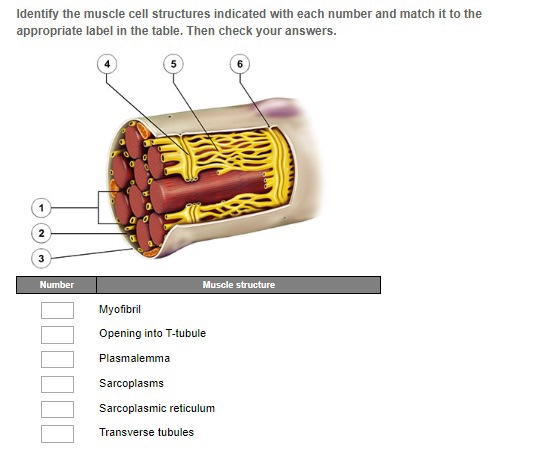
1. Myofibril
2. Sarcoplasma
3. Plasmalemma
4. Transverse tubules
5. Sarcoplasmic reticulum
6. Opening in to T tubule.
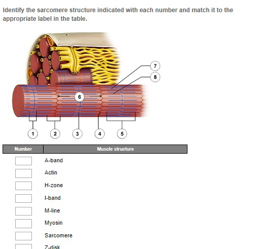
1. H - zone
2. I- band
3. M - line
4. Z- disk
5. A - band
6. Sarcomere
7. Actin
8. Myosin
Add Answer to:
Identify the muscle structure indicated with each number and match it to the appropriate label in...
Name Lab Partner 1. Part A Match the term with its correct description A. Sarcolemma B....
 Name Lab Partner 1. Part A Match the term with its correct description A. Sarcolemma B. Sarcomere C. Sarcoplasm D. Sarcoplasmic reticulum E. Endomysium F. Perimysium G. Epimysium H. Fascicle 1. Tendon Attaches muscle to bone Component of thick filament Component of thin filament Cytoplasm of a muscle cell Functional unit of a myofibril Cell membrane of a muscle cell Bundle of muscle fibers Produces ATP for muscle contraction Connective tissue that surrounds an individual muscle fiber Connective tissue that...
236 UNIT 12 introduction to Muscularsstern MusceTissue 2. Match each of the following terms with its...
 236 UNIT 12 introduction to Muscularsstern MusceTissue 2. Match each of the following terms with its description/function: transverse tubule l. connective tissue wrapping around a muscle fiber sarcoplasmic reticulum 2. organelle packed with myofilaments epimysium transverse tubule and its flanking terminalcisternae d. thin filament 4 invagination of the sarcolemma 5. connective tissue wrapping around fascicle myofibril 6. the region of sarcolemma across from the axon terminal f triad at the neuromuscular junction endomysium 7. consists of actin, troponin, and tropomyosin...
Label the image of a skeletal muscle fiber below. Actin Muscle fascicle Muscle fiber Muscle (organ)...
 Label the image of a skeletal muscle fiber below. Actin Muscle fascicle Muscle fiber Muscle (organ) Myosin Myofibril Sarcomere
Draw the basic structure of skeletal muscle. Label the following: Epimysium, Fascicle, Muscle Fibers, Perimysium, tendon....
 Draw the basic structure of skeletal muscle. Label the following: Epimysium, Fascicle, Muscle Fibers, Perimysium, tendon. 3. Define the following terms: 1. A Band 2. I Band 3. Z disk 4. H zone 5. M line Label the above on the following. Also include "sarcomere." 4. Complete the following chart Striated One or Size & Shape of Voluntary Special Or Non-striated Multiple Nuclei Cell Or Involuntary Features Muscle Type Cardiac Muscle Skeletal Muscle Smooth Muscle
PART A: Assessments match the terms in column A with the definitions in column B. Place...
 PART A: Assessments match the terms in column A with the definitions in column B. Place the letter of your choice in the space provided.12 Column A Column B a. Endomysium 1. Protein that comprises part of the thin filament with actin b. Epimysium 2. Cytoplasm of a muscle fiber c. Fascia d. Fascicle 3. Connective tissue located between adjacent muscles e. Myosin 4. Layer of connective tissue that separates a muscle into small bundles called f. Perimysium fascicles g....
SECTION 1: Structure of a Skeletal Muscle A. Layers of connective tissue separate a muscle into...
 SECTION 1: Structure of a Skeletal Muscle A. Layers of connective tissue separate a muscle into compartments. The layer called endomy- sium surrounds each muscle cell. Muscle cells are then bundled into fascicles, which are sur- rounded by perimysium. The entire muscle is composed of many fascicles, and is surround- ed by epimysium. All layers merge together to form the tendon that attaches the muscle to a bone. An outermost layer of connective tissue called fascia binds adjacent muscles together...
?Ex 17HW Core Lab Coaching Activity: Organization of Skeletal Muscles View Available Hint(s) Reset Help Z...
 ?Ex 17HW Core Lab Coaching Activity: Organization of Skeletal Muscles View Available Hint(s) Reset Help Z band or line myofibrils myosin T tubules sarcoplasmic reticulum troponin collagenous connective tissue layer surrounding the muscle. calcium storage site in skeletal muscles. rodlike structures that extend the length of the muscle fibers containing thin and thick filaments tubelike structures connecting the sarcolemma to the interior of the muscle fiber tendon : two terminal cisternae and a T tubule in the middle. triad tropomyosin...
Organization of Skeletal Muscles 17 A Matching Match each term in the left column with its...
 Organization of Skeletal Muscles 17 A Matching Match each term in the left column with its correct description from the light column 1. Sarcome A. banding patterns in muscle tissue 2. epimysium B storage site for calcium ions 3. perimyslum C. protein of thin filaments 1. endomysium D. group of muscle fibers 5. myofibril E. cylinder composed of filaments 6. Striations E protein of thick filaments 7. sarcolemma G. carries action potential deep into fiber 8. transverse tabule H. connective...
Multiple choice questions-Tor tes scantron questios s In a sarcomere, cross-bridge formation occurs specifically in the...
 Multiple choice questions-Tor tes scantron questios s In a sarcomere, cross-bridge formation occurs specifically in the 5. Skeletal muscle fibers are formed from embryonic cells calledwhich fuse together, making skeletal muscle cells multinucleated. 1. a. sarcomeres b. myofibrils c. myoblasts d. fascicles a. Z line. b. I band c. M line. d. H band e. zone of overlap. 6. Titin is a(n) a. elastic protein. b. thin filament protein. 2. A thin layer of connective tissue that surounds a muscle...
7. Use the key to label the structures on the thoracic region of the vertebral column....
 7. Use the key to label the structures on the thoracic region of the vertebral column. Key: a b. C. d. e intervertebral discs intervertebral foramina spinous prosesses thoracic vertebrae transverse processes Instructors may assign a portion of the Review Sheet questions using Mastering ABP EXERCISE REVIEW SHEET Microscopic Anatomy and Organization of Skeletal Muscle Name Lab Time Date Skeletal Muscle Cells and Their Organization into Muscles 1. Use the items in the key to correctly identify the structures described...
 Name Lab Partner 1. Part A Match the term with its correct description A. Sarcolemma B. Sarcomere C. Sarcoplasm D. Sarcoplasmic reticulum E. Endomysium F. Perimysium G. Epimysium H. Fascicle 1. Tendon Attaches muscle to bone Component of thick filament Component of thin filament Cytoplasm of a muscle cell Functional unit of a myofibril Cell membrane of a muscle cell Bundle of muscle fibers Produces ATP for muscle contraction Connective tissue that surrounds an individual muscle fiber Connective tissue that...
 236 UNIT 12 introduction to Muscularsstern MusceTissue 2. Match each of the following terms with its description/function: transverse tubule l. connective tissue wrapping around a muscle fiber sarcoplasmic reticulum 2. organelle packed with myofilaments epimysium transverse tubule and its flanking terminalcisternae d. thin filament 4 invagination of the sarcolemma 5. connective tissue wrapping around fascicle myofibril 6. the region of sarcolemma across from the axon terminal f triad at the neuromuscular junction endomysium 7. consists of actin, troponin, and tropomyosin...
 Label the image of a skeletal muscle fiber below. Actin Muscle fascicle Muscle fiber Muscle (organ) Myosin Myofibril Sarcomere
 Draw the basic structure of skeletal muscle. Label the following: Epimysium, Fascicle, Muscle Fibers, Perimysium, tendon. 3. Define the following terms: 1. A Band 2. I Band 3. Z disk 4. H zone 5. M line Label the above on the following. Also include "sarcomere." 4. Complete the following chart Striated One or Size & Shape of Voluntary Special Or Non-striated Multiple Nuclei Cell Or Involuntary Features Muscle Type Cardiac Muscle Skeletal Muscle Smooth Muscle
 PART A: Assessments match the terms in column A with the definitions in column B. Place the letter of your choice in the space provided.12 Column A Column B a. Endomysium 1. Protein that comprises part of the thin filament with actin b. Epimysium 2. Cytoplasm of a muscle fiber c. Fascia d. Fascicle 3. Connective tissue located between adjacent muscles e. Myosin 4. Layer of connective tissue that separates a muscle into small bundles called f. Perimysium fascicles g....
 SECTION 1: Structure of a Skeletal Muscle A. Layers of connective tissue separate a muscle into compartments. The layer called endomy- sium surrounds each muscle cell. Muscle cells are then bundled into fascicles, which are sur- rounded by perimysium. The entire muscle is composed of many fascicles, and is surround- ed by epimysium. All layers merge together to form the tendon that attaches the muscle to a bone. An outermost layer of connective tissue called fascia binds adjacent muscles together...
 ?Ex 17HW Core Lab Coaching Activity: Organization of Skeletal Muscles View Available Hint(s) Reset Help Z band or line myofibrils myosin T tubules sarcoplasmic reticulum troponin collagenous connective tissue layer surrounding the muscle. calcium storage site in skeletal muscles. rodlike structures that extend the length of the muscle fibers containing thin and thick filaments tubelike structures connecting the sarcolemma to the interior of the muscle fiber tendon : two terminal cisternae and a T tubule in the middle. triad tropomyosin...
 Organization of Skeletal Muscles 17 A Matching Match each term in the left column with its correct description from the light column 1. Sarcome A. banding patterns in muscle tissue 2. epimysium B storage site for calcium ions 3. perimyslum C. protein of thin filaments 1. endomysium D. group of muscle fibers 5. myofibril E. cylinder composed of filaments 6. Striations E protein of thick filaments 7. sarcolemma G. carries action potential deep into fiber 8. transverse tabule H. connective...
 Multiple choice questions-Tor tes scantron questios s In a sarcomere, cross-bridge formation occurs specifically in the 5. Skeletal muscle fibers are formed from embryonic cells calledwhich fuse together, making skeletal muscle cells multinucleated. 1. a. sarcomeres b. myofibrils c. myoblasts d. fascicles a. Z line. b. I band c. M line. d. H band e. zone of overlap. 6. Titin is a(n) a. elastic protein. b. thin filament protein. 2. A thin layer of connective tissue that surounds a muscle...
 7. Use the key to label the structures on the thoracic region of the vertebral column. Key: a b. C. d. e intervertebral discs intervertebral foramina spinous prosesses thoracic vertebrae transverse processes Instructors may assign a portion of the Review Sheet questions using Mastering ABP EXERCISE REVIEW SHEET Microscopic Anatomy and Organization of Skeletal Muscle Name Lab Time Date Skeletal Muscle Cells and Their Organization into Muscles 1. Use the items in the key to correctly identify the structures described...
Most questions answered within 3 hours.
-
A coach uses a new technique to train gymnasts. Seven
gymnasts were randomly selected and their...
asked 1 hour ago -
While rotating the tires on your car you notice a rock [mass =
0.1 Kg] stuck...
asked 3 hours ago -
Using MARS simulator, write MIPS programs according to
the following scenarios: Receive a positive integer number...
asked 5 hours ago -
An object in front of a concave mirror has a real image that is
11.5 cm...
asked 5 hours ago -
Consider the reaction, C3 H8 + O2 --> CO2 + H2O. How many
moles of O2...
asked 7 hours ago -
You and your opponent both roll a fair die. If you both roll the
same number,...
asked 7 hours ago -
In a study of the accuracy of fast food drive-through orders,
Restaurant A had 257 accurate...
asked 7 hours ago -
Identify and describe in detail the four categories of
institutions that could be included in a...
asked 7 hours ago -
In python
class Customer:
def __init__(self, customer_id, last_name, first_name, phone_number, address):
self._customer_id = int(customer_id)
self._last_name =...
asked 7 hours ago -
What is an example of a limitation in implementing a new
ERP system and how it...
asked 7 hours ago -
In a section of 9.7cm of an artery with a radius of 2.6mm there
is a...
asked 7 hours ago -
the two carboxylic acid groups of aspartic acid have different
acidities with pKa values of 2.1...
asked 7 hours ago