
Homework Answers
ANSWER :
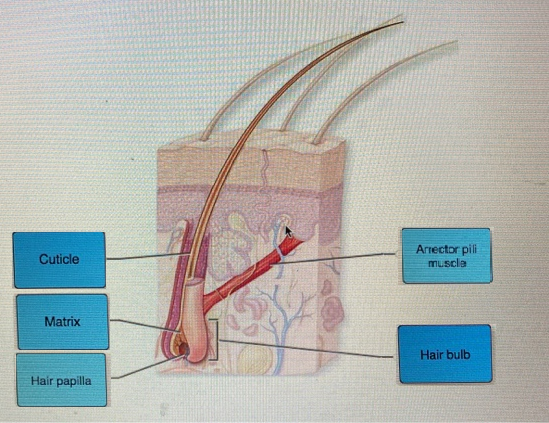
Labeling the hair structures that shown in the figure :-
Add Answer to:
Label the structures of hair in the figure. Hair bulb Cuticle Matrix Arrector pili muscle Hair...
10. Several structures of the hair are listed below. Identify each by matching its letter with...
 10. Several structures of the hair are listed below. Identify each by matching its letter with the appropriate area on the photomicrograph a cortex b. cuticle c. hair matrix d. hair papilla e medulla
Draw a longitudinal section of a hair follicle in the rectangular box to the right. Label...
 Draw a longitudinal section of a hair follicle in the rectangular box to the right. Label the following on your drawing: Glassy membrane • Hair shaft • Hair root Hair matrix Hair (dermal) papilla Melanocytes Internal root sheath External root sheath Medulla Cortex Cuticle Stop and think: What part of the Integumentary system do hair, nails and sweat glands derive from? Longitutinal section through a hair follicle
EXERCISE SIXThe 8-19: Match the stracture in colunn A with the coerect description in cokuan B a abe-shaped stractu...
 EXERCISE SIXThe 8-19: Match the stracture in colunn A with the coerect description in cokuan B a abe-shaped stracture that contains a haitr 9. Nail matrix b. visible portion of a nail sensory receptor that responds to deep pressare gland that is tmportant for regulating body temperature epidermal cell that produces melanin L part of a hair follicle that contains the hair matrix & glund that secretes a lipid substance called seburn h. cell type that comprises the majority of...
4 Identity and label the photograph of the skin slider Label epidermis, dermis Label arrector pill...
 4 Identity and label the photograph of the skin slider Label epidermis, dermis Label arrector pill muscle, hair follicle. hair, hair bulb, sebaceous gland, adipose tissue, sudoriferous gland JOJ 5. Identify and label the photograph of the skin model: dermis, stratum corneum, stratum granulosum, stratum spinosum, stratum basale
. For each true statement, write T. For each false statement, correct the underlined word(s) and...
 . For each true statement, write T. For each false statement, correct the underlined word(s) and insert your correction in the answer blank. 1. Greater amounts of the pigment sarotens are produced when the skin is exposed to the sun. 2. The most abundant protein in dead epidermal structures such as hair and nails is mclanin. 3. Sebum is an oily mixture of lipids, cholesterol, and cell fragments 1. The oldest epidermal cells in the epidermis are found in the...
Label the connective tissue structures associated with skeletal muscle. Answers will be used more than once....
 Label the connective tissue structures associated with skeletal muscle. Answers will be used more than once. Tendon Aponeurosis
8. The figure below is a diagrammatic representation of a small portion of a relaxed muscle...
 8. The figure below is a diagrammatic representation of a small portion of a relaxed muscle cell (bracket indicates the portion enlarged). (A) In the UPPER figure, LABEL the muscle cell / fiber and a myofibril. Then select different colors for the structures listed below and use them to COLOR the coding circles and corresponding structures on the lower figure. (B) In the LOWER figure, bracket and LABEL an A band, an l band, and a sarcomere. (C) Finally, DRAW...
Choc Muscle fiber nucleus Myofibrilot muscle fiber Figure 10.15. Details of the neuromuscular junction. Label the...
 Choc Muscle fiber nucleus Myofibrilot muscle fiber Figure 10.15. Details of the neuromuscular junction. Label the structures labeled with capital letters. The numbers 1-4 indicate the chain of events that occur at the neuromuscular junction. Put these events in chronological order by placing a l in the blank before the first event, etc. Refer back to Figure 10.15 for information. Acetylcholine diffuses across the synaptic cleft. Nerve impulse reaches the axon terminal. Synaptic vesicles release acetylcholine into the synaptic cleft....
2. Exercise 17-2 Using the microscope identify and label the cardiac muscle structures from figure 17.12...
 2. Exercise 17-2 Using the microscope identify and label the cardiac muscle structures from figure 17.12 (Page 462). Make a drawing or take a picture of your observations. 3. What is the name of the blood vessels that provides blood supply to the heart? From which artery does the blood vessels that provide blood to the heart come from? 4. During fetal life what structure allows blood flow from the right atrium to the left atrium? 5. What are the...
Using Figure 9.5 in your text, review muscle fiber structure Identify the labelled structures 1...
Using Figure 9.5 in your text, review muscle fiber structure Identify the labelled structures 1. 2. 3. 4. 5. 6. 2. Using the numbers from above identify the structure. Numbers can be used more than once _____ The structure that stores Ca+ _____ The structure that allows an action potential to move deep into the muscle fiber _____ The structure that has calcium release channels embedded in it _____ The muscle cell membrane _____ In-folds of the muscle cell membrane...
 10. Several structures of the hair are listed below. Identify each by matching its letter with the appropriate area on the photomicrograph a cortex b. cuticle c. hair matrix d. hair papilla e medulla
 Draw a longitudinal section of a hair follicle in the rectangular box to the right. Label the following on your drawing: Glassy membrane • Hair shaft • Hair root Hair matrix Hair (dermal) papilla Melanocytes Internal root sheath External root sheath Medulla Cortex Cuticle Stop and think: What part of the Integumentary system do hair, nails and sweat glands derive from? Longitutinal section through a hair follicle
 EXERCISE SIXThe 8-19: Match the stracture in colunn A with the coerect description in cokuan B a abe-shaped stracture that contains a haitr 9. Nail matrix b. visible portion of a nail sensory receptor that responds to deep pressare gland that is tmportant for regulating body temperature epidermal cell that produces melanin L part of a hair follicle that contains the hair matrix & glund that secretes a lipid substance called seburn h. cell type that comprises the majority of...
 4 Identity and label the photograph of the skin slider Label epidermis, dermis Label arrector pill muscle, hair follicle. hair, hair bulb, sebaceous gland, adipose tissue, sudoriferous gland JOJ 5. Identify and label the photograph of the skin model: dermis, stratum corneum, stratum granulosum, stratum spinosum, stratum basale
 . For each true statement, write T. For each false statement, correct the underlined word(s) and insert your correction in the answer blank. 1. Greater amounts of the pigment sarotens are produced when the skin is exposed to the sun. 2. The most abundant protein in dead epidermal structures such as hair and nails is mclanin. 3. Sebum is an oily mixture of lipids, cholesterol, and cell fragments 1. The oldest epidermal cells in the epidermis are found in the...
 Label the connective tissue structures associated with skeletal muscle. Answers will be used more than once. Tendon Aponeurosis
 8. The figure below is a diagrammatic representation of a small portion of a relaxed muscle cell (bracket indicates the portion enlarged). (A) In the UPPER figure, LABEL the muscle cell / fiber and a myofibril. Then select different colors for the structures listed below and use them to COLOR the coding circles and corresponding structures on the lower figure. (B) In the LOWER figure, bracket and LABEL an A band, an l band, and a sarcomere. (C) Finally, DRAW...
 Choc Muscle fiber nucleus Myofibrilot muscle fiber Figure 10.15. Details of the neuromuscular junction. Label the structures labeled with capital letters. The numbers 1-4 indicate the chain of events that occur at the neuromuscular junction. Put these events in chronological order by placing a l in the blank before the first event, etc. Refer back to Figure 10.15 for information. Acetylcholine diffuses across the synaptic cleft. Nerve impulse reaches the axon terminal. Synaptic vesicles release acetylcholine into the synaptic cleft....
 2. Exercise 17-2 Using the microscope identify and label the cardiac muscle structures from figure 17.12 (Page 462). Make a drawing or take a picture of your observations. 3. What is the name of the blood vessels that provides blood supply to the heart? From which artery does the blood vessels that provide blood to the heart come from? 4. During fetal life what structure allows blood flow from the right atrium to the left atrium? 5. What are the...
Most questions answered within 3 hours.
-
Under the influence of its drive force, a snowmobile is moving
at a constant velocity along...
asked 3 minutes ago -
What mechanisms Drive speciation??
(I.e. what was Dawins theory on the orgin of species, and how...
asked 1 hour ago -
The manager at a car assembly plant believes that the mean
assembly time for a car...
asked 2 hours ago -
Which of the following is true of electron capture?
A) It decreases the nuclide's mass number...
asked 3 hours ago -
Assuming an efficiency of 43.10%, calculate the actual yield of
magnesium nitrate formed from 114.9 g...
asked 4 hours ago -
The highly pathogenic bacterium Clostridium
perfringens causes gangrene, a disease that results in the
destruction of...
asked 6 hours ago -
In the context of situation analysis, which of the following is
a category for analysis in...
asked 6 hours ago -
In a study of the gas phase decomposition of sulfuryl chloride
at 600 K SO2Cl2(g)SO2(g) +...
asked 6 hours ago -
75 g of 2-propanol (C3H8O) and 25 g of pentane are mixed in a
200 mL...
asked 6 hours ago -
The 2800-turn coil in a dc motor has an area per turn of 1.1 ×
10-2...
asked 6 hours ago -
Draw a combinational logic circuit diagram with a symbol inside
the box for two I/P of...
asked 6 hours ago -
The cliché we use quite a lot in finance is: there is a need to
maximize...
asked 6 hours ago