
Homework Answers
enthalpically driven
Myoglobin is a monomeric protein that has 153 amino acids
residues. It consists of eight α-helicies connected through the
turns with an Oxygen binding site. It has a globular structure.
Myoglobin contains a heme (prosthetic) group which is responsible
for its main function (carrying of oxygen molecules to muscle
tissues). Myoglobin can exist in the oxygen free form,
deoxymyoglobin, or in a form in which the oxygen molecule is bound,
called oxymyoglobin. Myoglobin is a protein found in muscles that
binds oxygen with its heme group like hemoglobin. Heme group
consists of protoporphyrin organic component and an iron atom
located in its center. The heme group gives muscle and blood their
distinctive red color. The organic component consists of four
pyrrole rings that are linked by methine bridges. In addition, heme
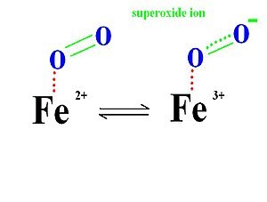
is responsible for the red color of the blood and muscle. Oxidation
of the iron atom (Fe2+ -> Fe3+) is mainly
responsible for the color of muscle and blood. At the center of
protporphyrin, the iron atom is bonded to nitrogen atoms from four
pyrrole rings. The iron atom can form two additional bonds, one on
each side of the heme plane. These binding sites are called the
fifth and sixth coordination sites. In myoglobin, the fifth
coordination site is occupied by the imidazole ring from a
histidine residue on the protein. This hisitidine is referred to as
the proximal histidine. The sixth coordination site is available to
bind oxygen. The iron atom in deoxymyoglobin lies about four
angstroms out of the plane of the protoporphyrin plane because it
is too big in that form to fit into the well defined hole.

The normal oxidation state of an iron atom has a positive two
charge (ferrous ion) instead of three charge (ferric ion) and it is
too large to fit into the plane of protoporphyrin. Thus, a ferrous
ion is often 0.4 Å away from the porphyrin plane. However, when
iron oxidized from ferrous ion (Fe2+) to ferric ion
(Fe3+), because the loss of one extra electron, forces
between protons and electrons increases so that the electron cloud
will penetrate more towards to the nucleus. As a result, the ferric
ion (Fe3+) has a smaller size then ferrous ion
(Fe2+) and fits into the protoporphyrin plane when it
attaches to an oxygen.
When oxygen leaves the myoglobin, it leaves as dioxygen rather than
superoxide. This is because superoxide can be damaging to many
biological process, and in the leaving of superdioxide, the iron
ion will be in the ferric state which stops biding oxygen.

The distal histidine amino acid from the hemoglobin protein
molecule further stabilizes the O2 molecule by
hydrogen-bonding interactions. 
Myoglobin is a protein molecule that has a similar structure and
function to hemoglobin. It is a smaller monomer of polypeptide
structure, a globular protein with amino acids and prosthetic heme
group binds to proximal histidine group while a distal histidine
group interact on the other side of the plane. It binds and stores
oxygen without concerning cooperativity. Most importantly, it is
the first protein structure to be studied.
Myoglobin follows the Michaelis-Menten Kinetic graph (as seen from the graph above). It follows the Michaelis-Menten kinetics because it is a simple chemical equilibrium.
FunctionEdit
The binding affinities for oxygen between myoglobin and hemoglobin are important factors for their function. Both myoglobin and hemoglobin binds oxygen well when the concentration of oxygen is really high (E.g. in Lung), however, hemoglobin is more likely to release oxygen in areas of low concentration (E.g. in tissues). Since hemoglobin binds oxygen less tightly than myoglobin in muscle tissues, it can effectively transport oxygen throughout the body and deliver it to the cells. Myoglobin, on the other hand, would not be as efficient in transferring oxygen. It does not show the cooperative binding of oxygen because it would take up oxygen and only release in extreme conditions. Myoglobin has a strong affinity for oxygen that allows it to store oxygen in muscle effectively. This is important when the body is starved for oxygen, such as during anaerobic exercise. During that time, carbon dioxide level in blood streams is extremely high and lactic acid concentration build up in muscles. Both of these factors cause myoglobin (and hemoglobins) to release oxygen, for protecting the body tissues from getting damaged under harsh conditions. If the concentration of myoglobin is high within the muscle cells, the organism is able to utilize the oxygen in its lungs for a much longer period of time.
Myoglobin, an iron-containing protein in muscle, receives oxygen from the red blood cells and transports it to the mitochondria of muscle cells, where the oxygen is used in cellular respiration to produce energy. Each myoglobin molecule has one heme prosthetic group located in the hydrophobic cleft in the protein. The function of myoglobin is notable from Millikan's review (1) in which he put together an accomplished study to establish that myoglobin is formed adaptively in tissues in response to oxygen needs and that myoglobin contributes to the oxygen supply of these tissues. Oxymyoglobin regulates both oxygen supply and utilization by acting as a scavenger of the bioactive molecule nitric oxide. Nitric oxide is generated continuously in the myocyte. Oxymyoglobin reacts with NO to form harmless nitrates, with concomitant formation of ferric myoglobin, which is recycled through the action of the intracellular enzyme metmyoglobin reductase. Flogel (2) conducted a study that showed how the interaction of NO and oxymyoglobin controls cardiac oxygen utilization.
Add Answer to:
Is 02 binding to myoglobin enthalpically or entropically driven? 。Enthalpically-driven Entropically-driven Question 6 Explain your answer...
5. Based on the binding curves of different versions of myoglobin shown below: -Myoglobin type 1...
 5. Based on the binding curves of different versions of myoglobin shown below: -Myoglobin type 1 -Myoglobin type 2 20 30 40 70 80 10g 50 60 PO, (torr) a. What are the pSos for the curves? (3 pts) b. Which type of Myoglobin binds oxygen better? Explain. (4 pts)
Answer all questions clearly and correctly Find an oxygen binding curve for myoglobin in your textbook....
 Answer all questions clearly and correctly
Find an oxygen binding curve for myoglobin in your textbook. How would you describe the shape of this 2. curve? 3. Explore the structure of hemoglobin using Proteopedia, Go to the Proteopedia site (http://www.proteopedia org/wiki/index.php/Main_Page) and search for hemoglobin. On the hemoglobin page, explore the structures and answer the tollowing questions. Note that you can rotate the hemoglobin molecule using your mouse and that clicking on green links changes the image of the structure....
Answer all questions clearly and correctly
Find an oxygen binding curve for myoglobin in your textbook. How would you describe the shape of this 2. curve? 3. Explore the structure of hemoglobin using Proteopedia, Go to the Proteopedia site (http://www.proteopedia org/wiki/index.php/Main_Page) and search for hemoglobin. On the hemoglobin page, explore the structures and answer the tollowing questions. Note that you can rotate the hemoglobin molecule using your mouse and that clicking on green links changes the image of the structure....
QUESTION 9 Which amino acid stabilizes ligand binding in the binding pocket of hemoglobin and myoglobin...
 QUESTION 9 Which amino acid stabilizes ligand binding in the binding pocket of hemoglobin and myoglobin via hydrogen bonding with the ligand? Distal Histidine Proximal Histidine Distal Valine Proximal phenylalanine QUESTION 10 Which of the following statement(s) is/are TRUE regarding enzymatic catalysis? Enzymes catalyze chemical transformations by altering the chemical equilibrium of the reactants and products. Enzymes catalyze chemical transformations by decreasing the free energy of the reactants. Enzymes catalyze chemical transformations by decreasing the free energy of the transition...
Write a paragraph of 3-5 sentences answering the following question. Your response should first answer the...
Write a paragraph of 3-5 sentences answering the following question. Your response should first answer the question and then go on to state physiological interpretation's behind your answer. Paragraphs that demonstrate complete and thorough reasoning will receive full points. Compared to hemoglobin, myoglobin has a lower binding affinity for O2 and a higher O2 binding capacity, two characteristics that allow myoglobin to efficiently provide skeletal muscle cells with a readily-accessible reservoir of O2.
please answer all. 14. [6 pts) Indicate which of the following statements is True (T) or...
 please answer all.
14. [6 pts) Indicate which of the following statements is True (T) or False (F) When water interacts with hydrophobic molecules, it becomes more ordered and entropy of the system increases The hydrophobic effect is predominant in protein stability The Ramachandran ploy shows combinations of dihedral angles in a polypeptide chain. A proteins function is directly related to the protein's secondary structure. The B sheet is primarily stabilized by R group interactions Clusters of secondary structures are...
please answer all.
14. [6 pts) Indicate which of the following statements is True (T) or False (F) When water interacts with hydrophobic molecules, it becomes more ordered and entropy of the system increases The hydrophobic effect is predominant in protein stability The Ramachandran ploy shows combinations of dihedral angles in a polypeptide chain. A proteins function is directly related to the protein's secondary structure. The B sheet is primarily stabilized by R group interactions Clusters of secondary structures are...
Question 6 (1 point) Relative to non-cooperative DNA binding, cooperative DNA binding of proteins leads to...
Question 6 (1 point) Relative to non-cooperative DNA binding, cooperative DNA binding of proteins leads to which of the following? Question 6 options: lower binding affinity increased binding to nonspecific sequences a sigmoidal binding curve a slower transition between unbound and completely bound protein as concentration increases tighter binding to nucleosomes
1. Predict and explain the trend in K+ binding constants (6 marks):
 1. Predict and explain the trend in K+ binding constants (6 marks):
Question 5 -a: For the shaft application in Question 2, the input shaft (2) is driven...
 Question 5 -a: For the shaft application in Question 2, the input shaft (2) is driven at a constant speed of 150 rev/min. Obtain a basic load rating for a ball bearing at A and B in shaft 1 for a life of 10 Khr with a combined reliability of R = 0.95 . Also select a proper 02-Series Deep-Groove ball bearing according to your calculations. Assume the distribution data from manufacturer 2, in table 11-6 in the textbook. 800...
please answer in details this biophysical chemistry question 5. The measured Hill coefficient for a binding...
 please answer in details this biophysical chemistry
question
5. The measured Hill coefficient for a binding to hemoglobin under Physiologie conditions, which has four binding site of for a, is only about 3. Why is this?
please answer in details this biophysical chemistry
question
5. The measured Hill coefficient for a binding to hemoglobin under Physiologie conditions, which has four binding site of for a, is only about 3. Why is this?
Enthalpically-favored, Entropically-unfavored Chemical Reaction Consider the chemical reaction A+B C+D, going in the indicated direction....
Enthalpically-favored, Entropically-unfavored Chemical Reaction Consider the chemical reaction A+B C+D, going in the indicated direction. Assume that it takes place under conditions of both constant temperature and constant pressure, and that the entropy of the products is less than the entropy of the reactants and the enthalpy of the products is less than the enthalpy of the reactants. (That is, both ΔS and ΔH are negative when the reaction goes in the indicated direction.) Answer the following question and...
 5. Based on the binding curves of different versions of myoglobin shown below: -Myoglobin type 1 -Myoglobin type 2 20 30 40 70 80 10g 50 60 PO, (torr) a. What are the pSos for the curves? (3 pts) b. Which type of Myoglobin binds oxygen better? Explain. (4 pts)
 Answer all questions clearly and correctly
Find an oxygen binding curve for myoglobin in your textbook. How would you describe the shape of this 2. curve? 3. Explore the structure of hemoglobin using Proteopedia, Go to the Proteopedia site (http://www.proteopedia org/wiki/index.php/Main_Page) and search for hemoglobin. On the hemoglobin page, explore the structures and answer the tollowing questions. Note that you can rotate the hemoglobin molecule using your mouse and that clicking on green links changes the image of the structure....
Answer all questions clearly and correctly
Find an oxygen binding curve for myoglobin in your textbook. How would you describe the shape of this 2. curve? 3. Explore the structure of hemoglobin using Proteopedia, Go to the Proteopedia site (http://www.proteopedia org/wiki/index.php/Main_Page) and search for hemoglobin. On the hemoglobin page, explore the structures and answer the tollowing questions. Note that you can rotate the hemoglobin molecule using your mouse and that clicking on green links changes the image of the structure....
 QUESTION 9 Which amino acid stabilizes ligand binding in the binding pocket of hemoglobin and myoglobin via hydrogen bonding with the ligand? Distal Histidine Proximal Histidine Distal Valine Proximal phenylalanine QUESTION 10 Which of the following statement(s) is/are TRUE regarding enzymatic catalysis? Enzymes catalyze chemical transformations by altering the chemical equilibrium of the reactants and products. Enzymes catalyze chemical transformations by decreasing the free energy of the reactants. Enzymes catalyze chemical transformations by decreasing the free energy of the transition...
 please answer all.
14. [6 pts) Indicate which of the following statements is True (T) or False (F) When water interacts with hydrophobic molecules, it becomes more ordered and entropy of the system increases The hydrophobic effect is predominant in protein stability The Ramachandran ploy shows combinations of dihedral angles in a polypeptide chain. A proteins function is directly related to the protein's secondary structure. The B sheet is primarily stabilized by R group interactions Clusters of secondary structures are...
please answer all.
14. [6 pts) Indicate which of the following statements is True (T) or False (F) When water interacts with hydrophobic molecules, it becomes more ordered and entropy of the system increases The hydrophobic effect is predominant in protein stability The Ramachandran ploy shows combinations of dihedral angles in a polypeptide chain. A proteins function is directly related to the protein's secondary structure. The B sheet is primarily stabilized by R group interactions Clusters of secondary structures are...
 1. Predict and explain the trend in K+ binding constants (6 marks):
 Question 5 -a: For the shaft application in Question 2, the input shaft (2) is driven at a constant speed of 150 rev/min. Obtain a basic load rating for a ball bearing at A and B in shaft 1 for a life of 10 Khr with a combined reliability of R = 0.95 . Also select a proper 02-Series Deep-Groove ball bearing according to your calculations. Assume the distribution data from manufacturer 2, in table 11-6 in the textbook. 800...
 please answer in details this biophysical chemistry
question
5. The measured Hill coefficient for a binding to hemoglobin under Physiologie conditions, which has four binding site of for a, is only about 3. Why is this?
please answer in details this biophysical chemistry
question
5. The measured Hill coefficient for a binding to hemoglobin under Physiologie conditions, which has four binding site of for a, is only about 3. Why is this?
Most questions answered within 3 hours.
-
What is the temperature of a 3.5L container with 1.502 mol
carbon dioxide at 10.76 atm?...
asked 5 minutes ago -
A dragster makes a pass down a track in exactly 7.03 seconds.
You do not know,...
asked 9 minutes ago -
1. Describe all
necessary steps and conditions for execution of a java".
2. What
is maven...
asked 10 minutes ago -
What evidence can you provide to illustrate why
crowdsourcing is able to reduce system development costs?...
asked 14 minutes ago -
If the value of the range is equal to zero, then this indicates
that:
A. there...
asked 15 minutes ago -
Hi, I was wondering if I could get some help on a java program
that I...
asked 20 minutes ago -
Given is the peptide Ala-Lys-Lys-Asp-Phe
1) what is the pI for this peptide? show your
calculations!...
asked 25 minutes ago -
Discuss the benefits of Inventory, Reservation,
& Menu Management Systems. Assuming you are the Manager for...
asked 29 minutes ago -
Which of the following is a concern of the Epic of
Gilgamesh?
human mortality
the destiny...
asked 30 minutes ago -
CONVERT THE FOLLOWING FUNCTION IN MARS SIMULATOR
(MIPS)
This FUNCTION should be Runnable on MARS (MIPS...
asked 35 minutes ago -
Week 8 forum SPHE461
The field of the Fitness Professional includes many
different titles, specialties, certifying...
asked 45 minutes ago -
1. In the short run, the marginal product of labor increases
then decreases because of what?...
asked 1 hour ago