
Homework Answers
A.
1). EcoRI produces fragments of sizes 2.9, 4.5, 6.2, 7.4, and 8.0 kbp..
HindIII: Cuts twice, so you will get a small piece and a large piece.. size of 2.8kbp
PstI- 1.4bp..
2). Is already answered by appliying formula:
%w/v = w×100/ v
B.
1).Watson Crick model says that
is a double-stranded, helical molecule. It consists of two sugar-phosphate backbones on the outside, held together by hydrogen bonds between pairs of nitrogenous bases on the inside.. And To cut DNA, all restriction enzymes make two incisions, once through each sugar-phosphate backbone (i.e. each strand) of the DNA double helix.
2).
There are two ways of gene transfer by which foreign DNA can be introduced into the host cells:
1. Indirect gene transfer: When the foreign DNA is inserted into host cells by the use of a vector, the method of gene transfer is called indirect gene transfer. Indirect gene transfer generally occurs through:
(i) Plant tumours: This method has been used successfully to transform plant cells, which are difficult to transform. The gene is first inserted into the Ti plasmid of soil bacterium Agrobacterium tumefaciens. This method employs the usage of tumour causing microbe Agrobacterium tumefaciens to transfer DNA, by inducing the tumour formation in plants.
(ii) Viruses: The vector is first incorporated into a virus, which is then used to infect cells, carrying the foreign gene along with its own genetic material.
2. Direct gene transfer: The method of gene transfer in which DA is inserted directly into host cell is called direct gene transfer. Direct gene transfer occurs through:
(i) Heat shock: Cells are incubated with vector in a solution containing calcium ions at 0°C. The temperature is then suddenly raised to about 40°C. This heat shock causes some of the cells to take up the vector, as sudden heat develops pores in the membrane.
(ii) Electroporation: Cells are subjected to high-voltage pulse, which temporarily disrupts the membrane and allows the vector to enter the cell.
(iii) Gene gun: This technique fires microscopic gold or tungsten particles coated with foreign DNA at cells using a compressed air gun (burst of helium).
(Iv) Microinjection: A cell is held on a glass pipette with diameter much smaller than the cell, under a microscope and foreign DNA is injected directly into the nucleus using an incredibly fine micropipette, which punctures the plasma membrane....
Role of restriction enzyme:-
To introduce foreign DNA into a circular vector, there are three-step process:
Cutting out the gene
first remove the gene of interest from the DNA sequences on either side of it. We use restriction enzymes to do the cutting. These enzymes, which came originally from bacteria, cut DNA at specific sites in the sequence. .
Opening up the vector
By using the same restriction enzymes as they used to cut out the gene in step 1. This turns the vector into a linear molecule and makes it ready to accept the new piece of DNA.
Sticking the vector and the gene together..
3) video is not given..
4). Restriction enzymes recognises specific base sequence is known as the recognition sequence ...
Each restriction endonuclease recognises a specific
palindromic nucleotide sequences in the DNA.
For example, the following sequences reads
the same on the two strands in 5' Æ 3' direction. This is also true
if
read in the 3' Æ 5' direction.
5' —— GAATTC —— 3'
3' —— CTTAAG —— 5'
Restriction enzymes cut the strand of DNA a little away from the
centre of the palindrome sites, but between the same two bases on
the opposite strands. This leaves single stranded portions at the
ends. There are
overhanging stretches called sticky ends on each strand ..eg
| EcoR1 | Escherichia coli |
5'GAATTC 3'CTTAAG |
5'---G AATTC---3' 3'---CTTAA G---5' |
| BamH1 | Bacillus amyloliquefaciens |
5'GGATCC 3'CCTAGG |
5'---G GATCC---3' 3'---CCTAG G---5' |
5).When cut by the same restriction enzyme, the resultant DNA fragments have the same kind of ‘sticky-ends’ and, these can be joined together (end-to-end) using DNA ligases ...
6). Purified pGLo DNA is cut by restriction enzymes .. If we know where the enzymes will cut the DNA, ywe can predict the DNA fragment sizes and we should see on the gel, and that will help us ascertain whether we have the correct plasmid.
We can also plot Restriction maps thatthat DNA maps that show restriction sites, the locations where specific restriction enzymes will cut the DNA.
Eg:- 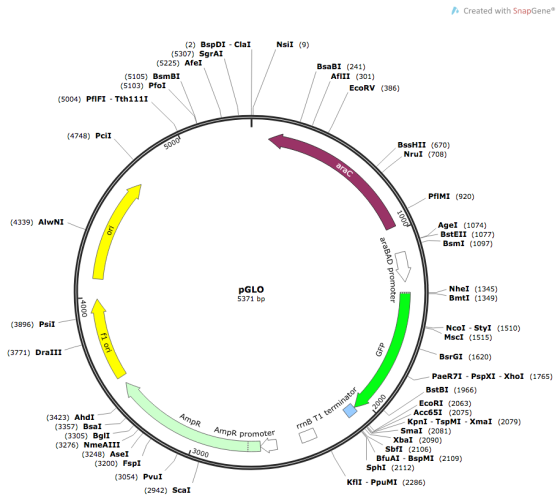
7). They are kept at same pH by TBE and TAE buffer...
Two important functions performed by gel electrophoresis buffer,
- It maintains the pH of reaction nearly neutral. The weak acid and base in buffer keep pH in the desired range.
- By maintaining neutral pH, it controls the net charge of molecules which helps in proper migration and separation of the molecule....
Add Answer to:
A. Your lab To see if you understand what you did in our lab, answer the...
please help if you can 3. You cloned a 20 kb piece of DNA, which contains...
 please help if you can
3. You cloned a 20 kb piece of DNA, which contains restriction sites as shown below. 281534 5 B 4 & 5 B = BamHl site, E = EcoRI site, H = Hindill site Numbers above the segments represent the sizes of the regions in kb. Draw and label (in the agarose gel below) the sizes of the fragments you would expect to see after complete digestion of this piece of DNA with the following...
please help if you can
3. You cloned a 20 kb piece of DNA, which contains restriction sites as shown below. 281534 5 B 4 & 5 B = BamHl site, E = EcoRI site, H = Hindill site Numbers above the segments represent the sizes of the regions in kb. Draw and label (in the agarose gel below) the sizes of the fragments you would expect to see after complete digestion of this piece of DNA with the following...
9. On Worksheet 16.IIIB is a restriction map of bacteriophage lambda. You digest some lambda DNA...
 9. On Worksheet 16.IIIB is a restriction map of bacteriophage lambda. You digest some lambda DNA with the enzymes BamHI and HindIII separately and then load the fragments into an agarose gel and perform electrophoresis. Next, you perform a Southern analysis using the 4,878-bp EcoRI lambda fragment as a probe. a. Draw a picture of the electrophoresis gel, using the outline of the stained electrophoresis gel in Worksheet 16.IIIB (the two smallest HindIII fragments will run off the gel.) b....
please help me with these questions Lab 8 Extension Activity: Plasmid Mapping and Restriction Enzymes Mapping...
 please help me with these questions
Lab 8 Extension Activity: Plasmid Mapping and Restriction Enzymes Mapping the Plasmid The first step in mapping a plasmid is to determine how many times a restriction site is found on that plasmid. Examine the results for plasmid 55 as an example. The data given in the following table are for the double digest using EcoRI and Pstl. Also, giving are the data for single digests by the individual enzymes. The numbers in the...
please help me with these questions
Lab 8 Extension Activity: Plasmid Mapping and Restriction Enzymes Mapping the Plasmid The first step in mapping a plasmid is to determine how many times a restriction site is found on that plasmid. Examine the results for plasmid 55 as an example. The data given in the following table are for the double digest using EcoRI and Pstl. Also, giving are the data for single digests by the individual enzymes. The numbers in the...
III. Subclone the gene into plasmid, extract the plasmid DNA. 5. You know that your insert...
 III. Subclone the gene into plasmid, extract the plasmid DNA. 5. You know that your insert (gene of interest, GOI) is flanked by the EcoRI sites, which makes this restriction enzyme a perfect candidate to cut out your gene. You also know that the GOI has a unique BamH1 restriction site. After subcloning the PCR product into the plasmid, a purified DNA preparation of the plasmid is digested to completion with BamHI restriction endonuclease. In separate reactions, the same preparation...
You have determined that a bacterial strain you are working with contains a single type of...
 You have determined that a bacterial strain you are working with
contains a single type of plasmid. You isolate the plasmid DNA and
digest separate portions of it with each of two different
restriction enzymes, BamHI and HpaI, and also perform a double
digest using both enzymes. You then fractionate the enzyme digests
on an agarose gel and stain the gel with ethidium bromideto
visualize the restriction fragment patterns. Your results are shown
below. Size markers (in nucleotide base pairs)...
You have determined that a bacterial strain you are working with
contains a single type of plasmid. You isolate the plasmid DNA and
digest separate portions of it with each of two different
restriction enzymes, BamHI and HpaI, and also perform a double
digest using both enzymes. You then fractionate the enzyme digests
on an agarose gel and stain the gel with ethidium bromideto
visualize the restriction fragment patterns. Your results are shown
below. Size markers (in nucleotide base pairs)...
Module 2 homework Laurel Jacqmai 0341 - Spring 20-MEEK > Activities and Due Dates > Module...
 Module 2 homework Laurel Jacqmai 0341 - Spring 20-MEEK > Activities and Due Dates > Module 2 homework Assignment Score: 20% Resources Hint Check Ans < Question 2 of 5 > EcoRI Control EcoRI BamHI BamHI Base pairs 10 kb Samples of a plasmid containing a segment of unknown DNA are digested using the restriction enzymes EcoRI, BamHI, and a combination of EcoRI and BamHI. The digests are then run on an agarose gel in order to separate the resulting...
The picture above represents an agarose gel that was used to analyze plasmid DNA after it...
 The picture above represents an agarose gel that was used to analyze plasmid DNA after it was cut with the restriction enzyme HindIll. The plasmid was incubated with Hindill until all of the available Hindlll cut sites were cut by HindIll. After running the sample on the gel, three bands were detected (Note that there are three wells shown at the top of the gel for loading samples, however, only the middle well was loaded with sample). Based on this...
B4. Answer ALL parts: The diagram below shows the restriction pattern of a plasmid cut with...
 B4. Answer ALL parts: The diagram below shows the restriction pattern of a plasmid cut with the enzymes BamHI, EcoRI and Ndel. The digests are carried out with each enzyme alone and then with different combinations of the three enzymes. Enzyme Fragment sizes kb BamHI 1.4 10.6 EcoRI 4.5 7.5 Ndel 2.5 9.5 BamHI + EcoRI 45 3.4 1.4 BamHI + Ndel 1.4 2.5 5.9 EcoRI + Ndel 0.5 2.0 2.5 7.0 (a) Draw an agarose gel with restriction fragments...
1. If a restriction enzyme cuts a circular plasmid twice, how many fragments would you see...
1. If a restriction enzyme cuts a circular plasmid twice, how many fragments would you see on the gel? 2. How would you estimate the total number of base pairs in a plasmid by looking at the DNA fragments of the digested plasmid on a gel? 3. If a linear 1kb DNA fragment has a restriction site that is located 50 bp from one end of the plasmid, what would you expect to see if the digested and undigested DNA...
help with questions 5 to 10 please PCB 3023L Lab #4 Protocol & Worksheet (30pt) You...
 help with questions 5 to 10 please
PCB 3023L Lab #4 Protocol & Worksheet (30pt) You may work in your lab groups durine class. but all written answers must be completed individually in your own words. 1) Using the plasmid map for orientation 1 and the cDNA map as a guide, complete the plasmid map for orientation #2. (4pt) 612 1318 1 - EcoRi EcoRI Xbal ECORV -Xbal- 1662 +Bell EcoRI EcoRV Not FP -- Xhol X 2015 PRSP +...
help with questions 5 to 10 please
PCB 3023L Lab #4 Protocol & Worksheet (30pt) You may work in your lab groups durine class. but all written answers must be completed individually in your own words. 1) Using the plasmid map for orientation 1 and the cDNA map as a guide, complete the plasmid map for orientation #2. (4pt) 612 1318 1 - EcoRi EcoRI Xbal ECORV -Xbal- 1662 +Bell EcoRI EcoRV Not FP -- Xhol X 2015 PRSP +...
 please help if you can
3. You cloned a 20 kb piece of DNA, which contains restriction sites as shown below. 281534 5 B 4 & 5 B = BamHl site, E = EcoRI site, H = Hindill site Numbers above the segments represent the sizes of the regions in kb. Draw and label (in the agarose gel below) the sizes of the fragments you would expect to see after complete digestion of this piece of DNA with the following...
please help if you can
3. You cloned a 20 kb piece of DNA, which contains restriction sites as shown below. 281534 5 B 4 & 5 B = BamHl site, E = EcoRI site, H = Hindill site Numbers above the segments represent the sizes of the regions in kb. Draw and label (in the agarose gel below) the sizes of the fragments you would expect to see after complete digestion of this piece of DNA with the following...
 9. On Worksheet 16.IIIB is a restriction map of bacteriophage lambda. You digest some lambda DNA with the enzymes BamHI and HindIII separately and then load the fragments into an agarose gel and perform electrophoresis. Next, you perform a Southern analysis using the 4,878-bp EcoRI lambda fragment as a probe. a. Draw a picture of the electrophoresis gel, using the outline of the stained electrophoresis gel in Worksheet 16.IIIB (the two smallest HindIII fragments will run off the gel.) b....
 please help me with these questions
Lab 8 Extension Activity: Plasmid Mapping and Restriction Enzymes Mapping the Plasmid The first step in mapping a plasmid is to determine how many times a restriction site is found on that plasmid. Examine the results for plasmid 55 as an example. The data given in the following table are for the double digest using EcoRI and Pstl. Also, giving are the data for single digests by the individual enzymes. The numbers in the...
please help me with these questions
Lab 8 Extension Activity: Plasmid Mapping and Restriction Enzymes Mapping the Plasmid The first step in mapping a plasmid is to determine how many times a restriction site is found on that plasmid. Examine the results for plasmid 55 as an example. The data given in the following table are for the double digest using EcoRI and Pstl. Also, giving are the data for single digests by the individual enzymes. The numbers in the...
 III. Subclone the gene into plasmid, extract the plasmid DNA. 5. You know that your insert (gene of interest, GOI) is flanked by the EcoRI sites, which makes this restriction enzyme a perfect candidate to cut out your gene. You also know that the GOI has a unique BamH1 restriction site. After subcloning the PCR product into the plasmid, a purified DNA preparation of the plasmid is digested to completion with BamHI restriction endonuclease. In separate reactions, the same preparation...
 You have determined that a bacterial strain you are working with
contains a single type of plasmid. You isolate the plasmid DNA and
digest separate portions of it with each of two different
restriction enzymes, BamHI and HpaI, and also perform a double
digest using both enzymes. You then fractionate the enzyme digests
on an agarose gel and stain the gel with ethidium bromideto
visualize the restriction fragment patterns. Your results are shown
below. Size markers (in nucleotide base pairs)...
You have determined that a bacterial strain you are working with
contains a single type of plasmid. You isolate the plasmid DNA and
digest separate portions of it with each of two different
restriction enzymes, BamHI and HpaI, and also perform a double
digest using both enzymes. You then fractionate the enzyme digests
on an agarose gel and stain the gel with ethidium bromideto
visualize the restriction fragment patterns. Your results are shown
below. Size markers (in nucleotide base pairs)...
 Module 2 homework Laurel Jacqmai 0341 - Spring 20-MEEK > Activities and Due Dates > Module 2 homework Assignment Score: 20% Resources Hint Check Ans < Question 2 of 5 > EcoRI Control EcoRI BamHI BamHI Base pairs 10 kb Samples of a plasmid containing a segment of unknown DNA are digested using the restriction enzymes EcoRI, BamHI, and a combination of EcoRI and BamHI. The digests are then run on an agarose gel in order to separate the resulting...
 The picture above represents an agarose gel that was used to analyze plasmid DNA after it was cut with the restriction enzyme HindIll. The plasmid was incubated with Hindill until all of the available Hindlll cut sites were cut by HindIll. After running the sample on the gel, three bands were detected (Note that there are three wells shown at the top of the gel for loading samples, however, only the middle well was loaded with sample). Based on this...
 B4. Answer ALL parts: The diagram below shows the restriction pattern of a plasmid cut with the enzymes BamHI, EcoRI and Ndel. The digests are carried out with each enzyme alone and then with different combinations of the three enzymes. Enzyme Fragment sizes kb BamHI 1.4 10.6 EcoRI 4.5 7.5 Ndel 2.5 9.5 BamHI + EcoRI 45 3.4 1.4 BamHI + Ndel 1.4 2.5 5.9 EcoRI + Ndel 0.5 2.0 2.5 7.0 (a) Draw an agarose gel with restriction fragments...
 help with questions 5 to 10 please
PCB 3023L Lab #4 Protocol & Worksheet (30pt) You may work in your lab groups durine class. but all written answers must be completed individually in your own words. 1) Using the plasmid map for orientation 1 and the cDNA map as a guide, complete the plasmid map for orientation #2. (4pt) 612 1318 1 - EcoRi EcoRI Xbal ECORV -Xbal- 1662 +Bell EcoRI EcoRV Not FP -- Xhol X 2015 PRSP +...
help with questions 5 to 10 please
PCB 3023L Lab #4 Protocol & Worksheet (30pt) You may work in your lab groups durine class. but all written answers must be completed individually in your own words. 1) Using the plasmid map for orientation 1 and the cDNA map as a guide, complete the plasmid map for orientation #2. (4pt) 612 1318 1 - EcoRi EcoRI Xbal ECORV -Xbal- 1662 +Bell EcoRI EcoRV Not FP -- Xhol X 2015 PRSP +...
Most questions answered within 3 hours.
-
Cisco packet tracer
Q1) Do you get any changes of IP address when packet is
traversing...
asked 11 minutes ago -
What is the pressure inside a 33.0 L container holding 106.4 kg
of argon gas at...
asked 59 minutes ago -
Question no 2
A housekeeping support department budgets its costs at
SR 40,000 per month plus...
asked 56 minutes ago -
A 1400Kg sports car accelerates from rest to 90km/h in 7.0s.
What is the average power...
asked 1 hour ago -
For the following reaction, 0.128 moles of
potassium hydrogen sulfateare mixed with
0.504 moles of potassium...
asked 5 hours ago -
1. What is the present value of $400, three years in the future
if the interest...
asked 5 hours ago -
The labor force minus the number of employed equals the number
of unemployed.
a. True
b....
asked 7 hours ago -
Determine the mass in units of grams [g] of 0.49 moles [mol]
of a new fictitious...
asked 8 hours ago -
A horizontal mass of M=5kg is on a spring and stretched to
x=0.5m when released from...
asked 9 hours ago -
26 of 50
"I have worked at the Arizona Humane Society for ten years, and
have...
asked 9 hours ago -
Compare and contrast zero based budgeting and incremental (or
base year) budgeting.
asked 9 hours ago -
4 pts 10. Which of the following hypothesis would be MOST
difficult to test experimentally? Group...
asked 9 hours ago