You digest a 10Kb Linear ECoRI dna fragment with TWO restriction enzymes and obtain the following...
You digest a 10Kb Linear ECoRI dna fragment with TWO restriction enzymes and obtain the following data Hind iii..... 3kb, 7kb BamHi ... 2 kb and 8kb HInd iii + bamHI ..... 1kb, 2kb, 7kb Draw a restriction map of this DNA fragment, labeling the sites for EcoRI, bahHI, and HIndiii
Homework Answers
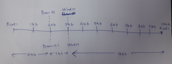
The above image will explain the map of the restriction site mentioned in the DNA fragment. Restriction enzymes are the enzymes which cut at specific nucleotide regions and has been useful in designing the map of DNA fragments.
Add Answer to:
You digest a 10Kb Linear ECoRI dna fragment with TWO restriction
enzymes and obtain the following...
I need help making a restriction digest map for both of these sets of fragments. Please...
 I need help making a
restriction digest map for both of these sets of fragments. Please
explain the steps and include drawing.
Draw restriction maps for the plasmids that produced the following fragments from restriction digestion: Digest Performed Size of DNA fragments produced BamHI BamHI/HindI 2.5kb & 1.5kb BamHI/EcoRI 3kb & 1kb HindIlI/EcoRI 3.5kb &0.5kb 4kp Digest Performed Size of DNA fragments produced BamHI BamHI & Sal1 4kb, 5kb BamHI & Pst1 6kb& 3kb Sal1&Pst1 1kb& 8kb kb
I need help making a
restriction digest map for both of these sets of fragments. Please
explain the steps and include drawing.
Draw restriction maps for the plasmids that produced the following fragments from restriction digestion: Digest Performed Size of DNA fragments produced BamHI BamHI/HindI 2.5kb & 1.5kb BamHI/EcoRI 3kb & 1kb HindIlI/EcoRI 3.5kb &0.5kb 4kp Digest Performed Size of DNA fragments produced BamHI BamHI & Sal1 4kb, 5kb BamHI & Pst1 6kb& 3kb Sal1&Pst1 1kb& 8kb kb
5. You have a linear DNA fragment and you wish to generate a restriction map using...
 5. You have a linear DNA fragment and you wish to generate a restriction map using Mstl and EcoR1. When the framgent is digested with Mst1 and the DNA is run on a gel you observe a 4kb and a 6kb fragment. When the DNA is digested with EcoR1, 1kb, 4kb and 5kb fragments are generated, A double digest using both enzymes produce 1kb, 2kb, 3kb and 4kb fragments. Explain these results and draw a restirction map consistent with these...
A 10 kb linear DNA molecule was digested with restriction enzyme EcoRI (E) and two fragments...
A 10 kb linear DNA molecule was digested with restriction enzyme EcoRI (E) and two fragments were produced of sizes 6 kb and 4 kb. The restriction enzyme HaeIII (H) also produced two fragments of sizes 9 kb and 1 kb. When the two enzymes HaeIII and EcoRI were used together, three fragments were produced of sizes 1 kb, 3 kb, and 6 kb. A 4 kb long cloned genomic probe from this region hybridized to the 3 kb and...
Restriction Mapping of a Linear DNA • 2 DNA is a linear and double-stranded DNA isolated...
 Restriction Mapping of a Linear DNA • 2 DNA is a linear and double-stranded DNA isolated from bacteriophage lambda. This bacteriophage is a virus that infects E. coli and can undergo specialized transduction (see the transduction lab lecture). Assuming you obtained the following fragments from the restriction digestion. Draw the restriction map for the EcoRI and Hindi II enzymes in the 2. DNA. Fragment Size EcoRI 15300 6200 5250 2100 EcoRI + HindIII 15300 4400 4000 2100 1800 Hind!!! 17100...
A linear fragment of DNA is cleaved with the individual restriction enzymes Hindill and Smal, and...
 A linear fragment of DNA is cleaved with the individual restriction enzymes Hindill and Smal, and then with a combination of the two enzymes. The fragments obtained are: Eco RV 2 kb, 3.5 kb, 9.5 kb Bam HI 6 kb, 9 kb Eco RV and Bam H12 kb, 3.5 kb, 4 kb. 5.5 kb Draw a restriction map of the DNA fragment. (5 marks). Model of example restriction map to show you how to give your answer: Eco R1 Hind...
Calculate the restriction map of plasmid JLB51given the following restriction digest data. HindIII 3.82 0.18 kb...
 Calculate the restriction map of plasmid JLB51given the following restriction digest data. HindIII 3.82 0.18 kb BamHI 2.35, 1.65 kb EcoRI 3.00, 1.0 kb HindIII, BamHI 2.35, 1.2, 0.27, 0.18 kb HindIII, EcoRI 1.87, 1.00, 0.95, 0.18 kb BamHI, EcoRI 1.6, 1.4, 0.75, 0.25 kb draw the circular map
A linear fragment of DNA is cleaved with the individual restriction enzymes Pst and Smal, and...
 A linear fragment of DNA is cleaved with the individual restriction enzymes Pst and Smal, and then with a combination of the two enzymes. The fragments obtained are: Pst! Sma! Pst and Smal 7 kb, 12 kb 2 kb, 8 kb, 9 kb 2 kb, 4 kb, 5 kb, 8 kb Draw a restriction map of the DNA fragment. (5 marks). Model of example restriction map to show you how to give your answer: Hind Ill kb 2 kb Eco...
9. On Worksheet 16.IIIB is a restriction map of bacteriophage lambda. You digest some lambda DNA...
 9. On Worksheet 16.IIIB is a restriction map of bacteriophage lambda. You digest some lambda DNA with the enzymes BamHI and HindIII separately and then load the fragments into an agarose gel and perform electrophoresis. Next, you perform a Southern analysis using the 4,878-bp EcoRI lambda fragment as a probe. a. Draw a picture of the electrophoresis gel, using the outline of the stained electrophoresis gel in Worksheet 16.IIIB (the two smallest HindIII fragments will run off the gel.) b....
A DNA molecule is cleaved with Hind III, Pst II, or the two enzymes together. The...
 A DNA molecule is cleaved with Hind III, Pst II, or the two enzymes together. The accompanying diagram shows the resulting electrophoresis gel, with band sizes indicated. Is the DNA molecule in question linear or circular? Linear circular Using the information in question 6. determine where the Pst 1 restriction site is located. Pstl cleave within the 7kb HindM fragment or within the 3kb HindM fragment'; 7kb fragment 3kb fragment neither fragment both fragments
You are using three restriction enzymes to digest a double-stranded DNA in which the sequence of...
You are using three restriction enzymes to digest a double-stranded DNA in which the sequence of the upper strand is 5'-TTGTCGATGCGAATTCGGTGATGGATCCTAGGTCGTGTAGCATGCATGCCGGATCCTAGCTGAGC'-3. The recognition sites of the enzymes are G'AATTC (EcoRI), G'GATCC (BamHI), and GCATG'C (SphI). The cleavage sites are indicated with '. Determine how long the DNA fragments will be after digesting the DNA with each of these enzymes individually. Additionally, determine the length of the fragments if you digest with both enzymes BamHI and SphI. In a drawing, show...
 I need help making a
restriction digest map for both of these sets of fragments. Please
explain the steps and include drawing.
Draw restriction maps for the plasmids that produced the following fragments from restriction digestion: Digest Performed Size of DNA fragments produced BamHI BamHI/HindI 2.5kb & 1.5kb BamHI/EcoRI 3kb & 1kb HindIlI/EcoRI 3.5kb &0.5kb 4kp Digest Performed Size of DNA fragments produced BamHI BamHI & Sal1 4kb, 5kb BamHI & Pst1 6kb& 3kb Sal1&Pst1 1kb& 8kb kb
I need help making a
restriction digest map for both of these sets of fragments. Please
explain the steps and include drawing.
Draw restriction maps for the plasmids that produced the following fragments from restriction digestion: Digest Performed Size of DNA fragments produced BamHI BamHI/HindI 2.5kb & 1.5kb BamHI/EcoRI 3kb & 1kb HindIlI/EcoRI 3.5kb &0.5kb 4kp Digest Performed Size of DNA fragments produced BamHI BamHI & Sal1 4kb, 5kb BamHI & Pst1 6kb& 3kb Sal1&Pst1 1kb& 8kb kb
 5. You have a linear DNA fragment and you wish to generate a restriction map using Mstl and EcoR1. When the framgent is digested with Mst1 and the DNA is run on a gel you observe a 4kb and a 6kb fragment. When the DNA is digested with EcoR1, 1kb, 4kb and 5kb fragments are generated, A double digest using both enzymes produce 1kb, 2kb, 3kb and 4kb fragments. Explain these results and draw a restirction map consistent with these...
 Restriction Mapping of a Linear DNA • 2 DNA is a linear and double-stranded DNA isolated from bacteriophage lambda. This bacteriophage is a virus that infects E. coli and can undergo specialized transduction (see the transduction lab lecture). Assuming you obtained the following fragments from the restriction digestion. Draw the restriction map for the EcoRI and Hindi II enzymes in the 2. DNA. Fragment Size EcoRI 15300 6200 5250 2100 EcoRI + HindIII 15300 4400 4000 2100 1800 Hind!!! 17100...
 A linear fragment of DNA is cleaved with the individual restriction enzymes Hindill and Smal, and then with a combination of the two enzymes. The fragments obtained are: Eco RV 2 kb, 3.5 kb, 9.5 kb Bam HI 6 kb, 9 kb Eco RV and Bam H12 kb, 3.5 kb, 4 kb. 5.5 kb Draw a restriction map of the DNA fragment. (5 marks). Model of example restriction map to show you how to give your answer: Eco R1 Hind...
 Calculate the restriction map of plasmid JLB51given the following restriction digest data. HindIII 3.82 0.18 kb BamHI 2.35, 1.65 kb EcoRI 3.00, 1.0 kb HindIII, BamHI 2.35, 1.2, 0.27, 0.18 kb HindIII, EcoRI 1.87, 1.00, 0.95, 0.18 kb BamHI, EcoRI 1.6, 1.4, 0.75, 0.25 kb draw the circular map
 A linear fragment of DNA is cleaved with the individual restriction enzymes Pst and Smal, and then with a combination of the two enzymes. The fragments obtained are: Pst! Sma! Pst and Smal 7 kb, 12 kb 2 kb, 8 kb, 9 kb 2 kb, 4 kb, 5 kb, 8 kb Draw a restriction map of the DNA fragment. (5 marks). Model of example restriction map to show you how to give your answer: Hind Ill kb 2 kb Eco...
 9. On Worksheet 16.IIIB is a restriction map of bacteriophage lambda. You digest some lambda DNA with the enzymes BamHI and HindIII separately and then load the fragments into an agarose gel and perform electrophoresis. Next, you perform a Southern analysis using the 4,878-bp EcoRI lambda fragment as a probe. a. Draw a picture of the electrophoresis gel, using the outline of the stained electrophoresis gel in Worksheet 16.IIIB (the two smallest HindIII fragments will run off the gel.) b....
 A DNA molecule is cleaved with Hind III, Pst II, or the two enzymes together. The accompanying diagram shows the resulting electrophoresis gel, with band sizes indicated. Is the DNA molecule in question linear or circular? Linear circular Using the information in question 6. determine where the Pst 1 restriction site is located. Pstl cleave within the 7kb HindM fragment or within the 3kb HindM fragment'; 7kb fragment 3kb fragment neither fragment both fragments
Most questions answered within 3 hours.
-
Hastings Entertainment has a beta of 0.64. If the market return
is expected to be 13.80...
asked 6 minutes ago -
9. Depository institutions are always:
a. illiquid
b. profitable
c. insolvent
d. all of the above...
asked 15 minutes ago -
Use AstroTurf Company's income statement below to answer the
following two questions. Answer these questions with...
asked 14 minutes ago -
How is a firm's task
environment different from its general environment? Provide
examples of both types...
asked 12 minutes ago -
What is one reason Innovators can adopt innovations so
early?
Group of answer choices
they are...
asked 15 minutes ago -
Show that min x^2, s.t. x>=2 has strong duality.
asked 15 minutes ago -
Using curved arrows show how the intermediate formed in this
reaction (Hexaphenylbenzene is prepared through a...
asked 21 minutes ago -
Two lightbulbs operate on the same current. Bulb A has four
times the power output of...
asked 17 minutes ago -
1. What five (5) basic parameters need to be measured
during a pump test in order...
asked 29 minutes ago -
One student ran a TLC of an unknown compound on a
silica gel plate and the...
asked 32 minutes ago -
Use inheritance to create a new class AudioRecording
based on Recording class that:
it will retain...
asked 37 minutes ago -
In the long run, an increase in the quantity of money ________
the value of money...
asked 43 minutes ago