


Question 4. Explain how Fig. 1A (arrow) result corroborates with the author hypothesis about Parkin aggresome and proteasome activity in PD?
Iarain Aeturmulation in Aggresomes Due to Proteasome Impairment* Received for publication, April 2, 2002, and in revised form, September 2 Published, JBC Papers in Press, October 2, 2002, DOI 10.1074jbe M20315 Eunsung Junn, Sang Seop Lee, Unsun T. Suhr, and M. Maral Mouradiant Prom the Genetic Pharmacology Unit, Esperimental Theropeutice Branch, NINDS, National Institutes of Health Bethesda, Maryland 20892-1406 Parkinson's disease (PD) is characterized by loss of dopaminergie neurons in the substantia nigra and by ally, overexpression of a-synuelein in transgenie models re- sults in the formation of intracellular protein aggregates and the presence of ubiquitinated cytoplasmie inelusions locomotor dysfunction (15, 16) known as Lewy bodies. a-Synuelein and Parkin are two Parkin, originally identified by positional cloning in families of the proteins associated with inherited forms of PD with autosomal recessive PD (13), is a ubiquitin-protein isopep- and are found in Lewy bodies. Whereas numerous re- tide ligase (E3) (17, 18). This 465-amino acid protein has mild ports indicate the tendeney of a-synuclein to aggregate homology to ubiquitin at its N terminus and contains two RING both in vitro and in vivo, no information is available finger domains at its C terminus. Parkin exerts its ubiquitin about similar physical properties for Parkin. Here we ligase function through interactions between its RING finger show that proteasome inhibitors leads to the formation of aggre- some-like perinuclear inclusions. These eosinophilic in- xpression of Parkin in the presence of domain and E2-conjugating enzymes. In addition to ubiquiti- clusions share many characteristies with Lewy bodies, osylated a-synuclein, including a core and halo organization, immunoreactiv- kin ubiquitinates itself as an early step in its proteasome- ity to ubiquitin, a-synuclein, synphilin-1, Parkin, molec- mediated degradation (18, 21) ular chaperones, and proteasome subunit as well as To date, several studies (22, 23) have addressed the tendency staining of some with thioflavin S. We propose that the of a-synuclein to aggregate as ubiquitinated inclusions, but no process of Lewy body formation may be akin to that of information is available about the ability of Parkin to aggre- aggresome-like structures. The tendeney of wild-type gate. In this report, we show that overexpression of Parkin in Parkin to aggregate and form inclusions may have im- the presence of a proteasome inhibitor leads to the accumula plications for the pathogenesis of sporadic PD tion of Parkin aggregates as single, large, eosinophilic peri- nuclear inclusions consisting of a core and a halo. These struc- tures are similar to aggresomes (24), the formation of which
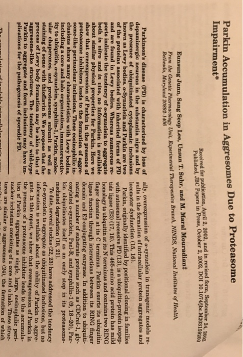
MG-132 t HMW 220- 127 84- 41ー
FIG. 1. Parkin forms ubiquitin-positive insoluble HMW com- plexes. A, HEK 293T cells transiently transfected with FLAG-Parkin were divided into two dishes and were either untreated or treated with 5 MG-132 for 16 h. Cells were then lysed in a buffer containing 1% Triton X-100 and fractionated into soluble (S) and insoluble (D) frac- tions, followed by Western blotting using anti-FLAG (M2) antibody. Filled arrow points to monomeric Parkin. B, Triton X-100-insoluble fraction from HEK 293T cells transfected with FLAG-Parkin were either untreated (- or treated (+) with 20 mM DTT prior to Western blot analysis. Filled arrow indicates monomeric Parkin. C, Tritorn X-100-insoluble fraction was subjected to immunoprecipitation with anti-FLAG (M2) antibody and followed by Western blot with anti- ubiquitin (P4D1) antibody. IP indicates the immunoprecipitated sam- ple, and T indicates total protein prior to immunoprecipitation. CTTAATGCAGATCTTCGTGAAGACTCTG-3' and 5'-GGCGAATTCT ACCCACCCTGAGACGGAGTAC-3'. The amplified sequence was in- serted into pHM6 (Roche Molecular Biochemicals) to express HA- tagged ubiquitin. Cell Culture and Transfection-cOS-7 and human embryonic kidney HEK 293T cell lines were maintained in Dulbecco's modified Eagle's medium containing 10% fetal bovine serum, PC12 cells were cultured in Dulbecco's modified Eagle's medium containing 10% horse serum and 5% fetal bovine serum. Transfections were performed using FuGENE 6 reagent (Roche Molecular Biochemicals) according to the supplier's instructions. Cells were cultured for 24 h after transfection Immunoprecipitation and Western Blot-Cells were lysed in a buffer containingPBSwith 1% Triton X-100 and a mixture of protease inhib- itors (Roche Molecular Biochemicals). After homogenizing with 20 strokes using a Dounce homogenizer, cells were centrifuged at 100,000 × g at 4 °C for 30 min. The soluble and insoluble fractions were -
Homework Answers
Parkin is a protein-ubiquitin E3 ligase connected to Parkinson's sickness. Albeit a few substrates of parkin have been distinguished, the subcellular area for parkin to perceive and ubiquitinate its objectives is misty. Here we report that parkin was collected in the centrosome when SH-SY5Y or transfected HEK293 cells were treated with the proteasome inhibitor lactacystin. The particular enlistment of parkin was subject to fixation and length of the treatment, and was joined by the centrosomal gathering of ubiquitinated proteins and CDCrel-1, a substrate of parkin. The enrollment of parkin was evidently interceded through its official to γ-tubulin, which has been appeared to aggregate in the centrosome because of misfolded proteins. Moreover, the impact was repealed by the microtubule-depolymerizing drug colchicine or the microtubule-settling drug taxol, which demonstrates that the unblemished microtubule arrange is required for the centrosomal enrollment of parkin. Taken together, our information recommend that the lactacystin-instigated aggregation of parkin in the centrosome assumes a critical job in the ubiquitination of misfolded substrates gathered there. This procedure may give a subcellular region to parkin to ubiquitinate and corrupt protein totals basically engaged with the pathogenesis of Parkinson's malady.
Add Answer to:
Q 4. Explain how Fig.1A (arrow) result corroborates with the author hypothesis about Parkin aggre...
Parkin Mannosidase II Merge Parkin Y-Adaptin Merge Parkin BIP Merge Parkin LAMP-1 Merge Parkin Ubiquitin Merge...
 Parkin Mannosidase II Merge Parkin Y-Adaptin Merge Parkin BIP Merge Parkin LAMP-1 Merge Parkin Ubiquitin Merge FIG. 2. Immunocytochemical detection of overexpressed Par- kin. COS-7 cells were transfected with FLAG-Parkin and subjected to co-immunostaining with anti-FLAG antibody (green) along with the Golgi complex marker mannosidase II (A), trans-Golgi network marker yadaptin (B), endoplasmic reticulum marker BIP/GRP78 (C), and ly- sosomal marker LAMP-1 (D), all with red fluorescence. E, COS-7 cells transfected with FLAG-Parkin were immunostained with anti-FLAG antibody (green) and...
 Parkin Mannosidase II Merge Parkin Y-Adaptin Merge Parkin BIP Merge Parkin LAMP-1 Merge Parkin Ubiquitin Merge FIG. 2. Immunocytochemical detection of overexpressed Par- kin. COS-7 cells were transfected with FLAG-Parkin and subjected to co-immunostaining with anti-FLAG antibody (green) along with the Golgi complex marker mannosidase II (A), trans-Golgi network marker yadaptin (B), endoplasmic reticulum marker BIP/GRP78 (C), and ly- sosomal marker LAMP-1 (D), all with red fluorescence. E, COS-7 cells transfected with FLAG-Parkin were immunostained with anti-FLAG antibody (green) and...
Most questions answered within 3 hours.
-
A circular coil of radius 0.120 m contains a single turn and is
located in a...
asked 4 minutes ago -
help me out
Velocity v = גf, wavelength ג = v/f and
Relative frequency = frequency...
asked 12 minutes ago -
1) Adrenaline is the hormone that triggers the
release of extra glucose molecules in times of...
asked 7 minutes ago -
In a competitive inhibition, Km is increased while Vmax is
unchanged. An enzyme is being assayed...
asked 47 minutes ago -
Applied manufacturing overheads are overheads allocated to the
production process according to a tariff, based on...
asked 44 minutes ago -
you are required to develop a simple HR application for a small
accounting firm that wishes...
asked 44 minutes ago -
The following present value factors are provided for use in this
problem.
Periods
Present Value
of...
asked 49 minutes ago -
Celine beauty is a new cosmetics manufacturer that produces
skincare box and makeup box. Demand over...
asked 56 minutes ago -
Eleanor and Kyoko are roommates. They spend most of their time
studying (of course), but they...
asked 1 hour ago -
Answer the questions
1.What is the value of a stock based on the dividend-growth
model if...
asked 1 hour ago -
Per the Barron's report the average weeks unemployed is 21.5
(population mean) with a population standard...
asked 1 hour ago -
1.. All following elements have been identified as
important to supporting school’s level of involvement with...
asked 1 hour ago