3. In the photomicrograph of dividing root cells in the margin, identify interphase and the following...

![REVIEWING YOUR KNOWLEDGE 1. Define the examples mitosis, m centrosom complex, furrow, d. 2. Describe DOLOed 3. In the] interp](http://img.homeworklib.com/questions/c6844470-e099-11eb-bffd-c5e551cb75d2.png?x-oss-process=image/resize,w_560)
Homework Answers
Answer:
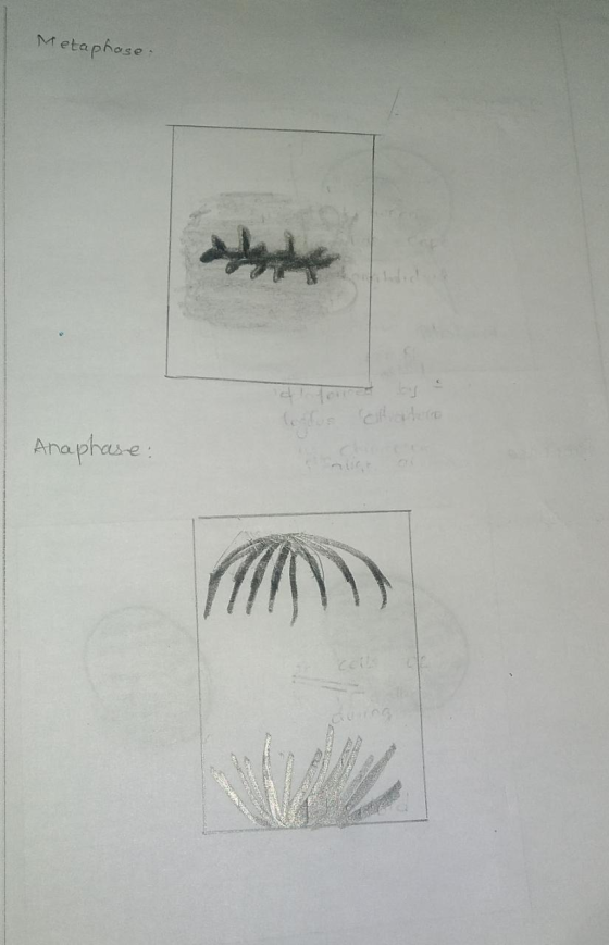
Hi, please find your answer in handwritten pictures (5)


 I love’d to hear your feedback
and Rating. If you like the answer please give me a positive
rating, which will boost me more. Thank you.
I love’d to hear your feedback
and Rating. If you like the answer please give me a positive
rating, which will boost me more. Thank you.
Add Answer to:
3. In the photomicrograph of dividing root cells in the margin, identify interphase and the following...
Click "View Image" to see a diagram of meiosis and mitosis. How many daughter cells are...
 Click "View Image" to see a diagram of meiosis and mitosis. How many daughter cells are produced by each of these processes? a) 46 for mitosis; 4 for meiosis b) 4 for mitosis; 2 for meiosis c) 2 for mitosis; 2 for meiosis d) 2 for mitosis; 4 for meiosis Comparison Meiosis - Mitosis MITOSIS MEIOSIS Interphase Interphase Prophase 1 Prometaphase Metaphase 1 Meiosis Anaphase 1 Telophase 1 Cytokinesis Prophase II Prophase Prometaphase Prometaph. I Metaphase Metaph. II 1200eli Meiosis...
1 bleul variation is accomplished. Crossing over-Does it occur? If so, name the stage it occurs....
 1 bleul variation is accomplished. Crossing over-Does it occur? If so, name the stage it occurs. 2. Mitosis is best described as a. cell division of reproductive cells. b. cell division of somatic cells into two diploid daughter cells. c. unequal division of the cytoplasm. d. cell division resulting in 4 haploid cells. Matching: Match phases of the mitotic cycle with characteristic event. 3. Interphase a. formation of cleavage furrow or cell plate 4. Prophase b. chromosomes line up along...
Examine the onion root tip slide images on the following pages. There are four images, each...
Examine the onion root tip slide images on the following pages. There are four images, each displaying a different field of view. Pick one of the images, and count the number of cells in each stage. Then count the total number of cells in the image. Record the image you selected and counts in Table 2. Calculate the time spent by a cell in each stage based on the 24 hour cycle: Hours of Stage = 24 x Number of...
What is the diploid number of chromosomes in this plant? _______________ ["2N=4", "N=8", "2N=16", "2N=8", "N=4",...
 What is the diploid number of chromosomes in this plant?
_______________ ["2N=4", "N=8", "2N=16", "2N=8", "N=4", "N=16"]
Give the names of each stage of mitosis or meiosis shown in
the:
left panel:
________________ ["anaphase of meiosis I", "metaphase
of meiosis I", "prophase of meiosis II", "metaphase of mitosis",
"telophase of mitosis", "anaphase of meiosis II", "anaphase of
mitosis", "prophase of mitosis", "telophase of meiosis II",
"metaphase of meiosis II", "telophase of meiosis I", "prophase of
meiosis...
What is the diploid number of chromosomes in this plant?
_______________ ["2N=4", "N=8", "2N=16", "2N=8", "N=4", "N=16"]
Give the names of each stage of mitosis or meiosis shown in
the:
left panel:
________________ ["anaphase of meiosis I", "metaphase
of meiosis I", "prophase of meiosis II", "metaphase of mitosis",
"telophase of mitosis", "anaphase of meiosis II", "anaphase of
mitosis", "prophase of mitosis", "telophase of meiosis II",
"metaphase of meiosis II", "telophase of meiosis I", "prophase of
meiosis...
Table 2: Mitosis Data Number of Cells in Stage Total Number of Cells Stage Calculated %...
 Table 2: Mitosis Data Number of Cells in Stage Total Number of Cells Stage Calculated % of Time Spent in Each Stage Interphase Prophase Metaphase Anaphase Telophase Cytokinesis
Question 1: The cell cycle has these stages
Question 1: The cell cycle has these stages A. GI, S, G2, M B. GI, S, BI, CL C. GI, S, G2, CI D. S, G2, NI, MI Question 2: During interphase, the following occurs A. Sister chromatids separate. B. Chromosome duplication takes place. C. Chromatin becomes tightly coiled. D. The mitotic spindle forms. Question 3: The correct sequence of stages of mitosis is A. prophase, metaphase, anaphase, telophase B. telophase, prophase, interphase, anaphase, metaphase C. interphase, anaphase, prophase, metaphase, telophase B. metaphase, prophase, anaphase, telophase Question 4: Human cells contain 23 pairs of chromosomes A....
Suppose that in an experiment you observed 113 cells in interphase, 24 cells in prophase, 9...
 Suppose that in an experiment you observed 113 cells in
interphase, 24 cells in prophase, 9 cells in metaphase, 12 cells in
anaphase, and 3 cells in telophase stages. If the complete cell
cycle (i.e. G1+S+G2+mitosis; see Figure 1-2 of the Lab Manual)
requires 24 hours, calculate the average duration of each stage in
the cycle. Show all calculations.
QUESTION 1 OF 10 Please read Chapter 1 of the Lab Manual and the relevant Power Point lecture in detail, and...
Suppose that in an experiment you observed 113 cells in
interphase, 24 cells in prophase, 9 cells in metaphase, 12 cells in
anaphase, and 3 cells in telophase stages. If the complete cell
cycle (i.e. G1+S+G2+mitosis; see Figure 1-2 of the Lab Manual)
requires 24 hours, calculate the average duration of each stage in
the cycle. Show all calculations.
QUESTION 1 OF 10 Please read Chapter 1 of the Lab Manual and the relevant Power Point lecture in detail, and...
Interphase Melosis 1 Draw in the chromosomes for each stage of Meiosis. Your cell has a...
 Interphase Melosis 1 Draw in the chromosomes for each stage of Meiosis. Your cell has a diplod # of 6 (haploid # of 3). Use two contrasting colors for your homologous chromosome pair. Prophase 1 Metaphase 1 Anaphase 1 Telophase 1 Melosis 2 Prophase 2 Metaphase 2 Anaphase 2 Telophase 2 4 daughter cells
1. If the amount of DNA in a somatic cell equals C during G1 of interphase,...
 1. If the amount of DNA in a somatic cell equals C during G1 of interphase, how much DNA is present in the cell during each phase of mitosis and meiosis? (2pts) Anaphase Telophase Cytokinesis Prophase 20 Metaphase T Mitosis Meiosis 1 Meiosis 2 2 Define the three things that are responsible for genetic variation and explain how they lead to genetic variation (1.5pts)
5. For this part of the lab you will be observing a whitefish blastula under the...
5. For this part of the lab you will be observing a whitefish blastula under the microscope. A blastula is a developmental stage in many animals. It occurs shortly after fertilization and the formation of the zygote. It is the “ball of cells” stage of development, where cells are dividing rapidly to form the multicellular organism. Note: In this simulation early and late prophase are combined into one stage: prophase. 1. Go to the Virtual Microscope (Links to an external...
 Click "View Image" to see a diagram of meiosis and mitosis. How many daughter cells are produced by each of these processes? a) 46 for mitosis; 4 for meiosis b) 4 for mitosis; 2 for meiosis c) 2 for mitosis; 2 for meiosis d) 2 for mitosis; 4 for meiosis Comparison Meiosis - Mitosis MITOSIS MEIOSIS Interphase Interphase Prophase 1 Prometaphase Metaphase 1 Meiosis Anaphase 1 Telophase 1 Cytokinesis Prophase II Prophase Prometaphase Prometaph. I Metaphase Metaph. II 1200eli Meiosis...
 1 bleul variation is accomplished. Crossing over-Does it occur? If so, name the stage it occurs. 2. Mitosis is best described as a. cell division of reproductive cells. b. cell division of somatic cells into two diploid daughter cells. c. unequal division of the cytoplasm. d. cell division resulting in 4 haploid cells. Matching: Match phases of the mitotic cycle with characteristic event. 3. Interphase a. formation of cleavage furrow or cell plate 4. Prophase b. chromosomes line up along...
 What is the diploid number of chromosomes in this plant?
_______________ ["2N=4", "N=8", "2N=16", "2N=8", "N=4", "N=16"]
Give the names of each stage of mitosis or meiosis shown in
the:
left panel:
________________ ["anaphase of meiosis I", "metaphase
of meiosis I", "prophase of meiosis II", "metaphase of mitosis",
"telophase of mitosis", "anaphase of meiosis II", "anaphase of
mitosis", "prophase of mitosis", "telophase of meiosis II",
"metaphase of meiosis II", "telophase of meiosis I", "prophase of
meiosis...
What is the diploid number of chromosomes in this plant?
_______________ ["2N=4", "N=8", "2N=16", "2N=8", "N=4", "N=16"]
Give the names of each stage of mitosis or meiosis shown in
the:
left panel:
________________ ["anaphase of meiosis I", "metaphase
of meiosis I", "prophase of meiosis II", "metaphase of mitosis",
"telophase of mitosis", "anaphase of meiosis II", "anaphase of
mitosis", "prophase of mitosis", "telophase of meiosis II",
"metaphase of meiosis II", "telophase of meiosis I", "prophase of
meiosis...
 Table 2: Mitosis Data Number of Cells in Stage Total Number of Cells Stage Calculated % of Time Spent in Each Stage Interphase Prophase Metaphase Anaphase Telophase Cytokinesis
 Suppose that in an experiment you observed 113 cells in
interphase, 24 cells in prophase, 9 cells in metaphase, 12 cells in
anaphase, and 3 cells in telophase stages. If the complete cell
cycle (i.e. G1+S+G2+mitosis; see Figure 1-2 of the Lab Manual)
requires 24 hours, calculate the average duration of each stage in
the cycle. Show all calculations.
QUESTION 1 OF 10 Please read Chapter 1 of the Lab Manual and the relevant Power Point lecture in detail, and...
Suppose that in an experiment you observed 113 cells in
interphase, 24 cells in prophase, 9 cells in metaphase, 12 cells in
anaphase, and 3 cells in telophase stages. If the complete cell
cycle (i.e. G1+S+G2+mitosis; see Figure 1-2 of the Lab Manual)
requires 24 hours, calculate the average duration of each stage in
the cycle. Show all calculations.
QUESTION 1 OF 10 Please read Chapter 1 of the Lab Manual and the relevant Power Point lecture in detail, and...
 Interphase Melosis 1 Draw in the chromosomes for each stage of Meiosis. Your cell has a diplod # of 6 (haploid # of 3). Use two contrasting colors for your homologous chromosome pair. Prophase 1 Metaphase 1 Anaphase 1 Telophase 1 Melosis 2 Prophase 2 Metaphase 2 Anaphase 2 Telophase 2 4 daughter cells
 1. If the amount of DNA in a somatic cell equals C during G1 of interphase, how much DNA is present in the cell during each phase of mitosis and meiosis? (2pts) Anaphase Telophase Cytokinesis Prophase 20 Metaphase T Mitosis Meiosis 1 Meiosis 2 2 Define the three things that are responsible for genetic variation and explain how they lead to genetic variation (1.5pts)
Most questions answered within 3 hours.
-
If 20 g of Na2SO4 is reacted with 20 g of
Al(NO3)3 according to the following...
asked 20 seconds ago -
Using the OSHA web site find the OSHA regulation for personal eye
protection. Write a summary...
asked 4 minutes ago -
In 600 words, answer the following
1) Why has the transfer of defense technologies to domestic...
asked 4 minutes ago -
One difference between periodic and perpetual inventory systems
is:
Multiple Choice Cost of goods sold is...
asked 6 minutes ago -
Balance the following oxidation-reduction equations using redox
methods:
Cu + H+ --------> Cu+ +
H2
asked 25 minutes ago -
For a voltaic cell based on the reaction below, which statement
is correct?
Zn(s)+2H+(aq)→Zn2+(aq)+H2(g)
Zn2+(aq) is...
asked 9 minutes ago -
For the balanced reaction: CaCl2 (aq) + Na2CO3 (aq) -> CaCO3
(s) + 2 NaCl (aq),...
asked 19 minutes ago -
1. If ln(x)=ln(x)= -0.2 , what does xx equal? Round
your answer to three significant figures. The...
asked 15 minutes ago -
what is the current research being done on the hexokinase
enzyme?
asked 21 minutes ago -
An incline weighing 1,106 kg with its passengers travels uphill
800 meters on a 30 degree...
asked 22 minutes ago -
Let’s assume you want to retire with $1,000,000 in your
investment portfolio. Given that your investment...
asked 22 minutes ago -
profit motivation is one of the biggest differences between
public and private organizations. what about resource...
asked 21 minutes ago